Figure 1. Conidia of Fusicoccum putrefaciens on PDA (400x).According to symptoms upright dieback, blossom and ovary blight and end rot were caused byFusicoccum putrefaciens Shear. The teleomorph stage caused by Godronia cassandrae Peck f.vaccinii Groves was not found in Latvia. The causal agent of the disease was identified based onsymptoms and the morphological characteristics described by Гopлeнкo et al., 1996 and Caruso F.L., 1995.Phomopsis vaccinii from upright dieback, blossom and ovary blight and viscid rot samples wasdetected. Uprights, blossoms and ovaries turned brown and died, but viscid rot was off-color,slightly mottled or yellowish brown and firm but wet inside with a viscous, sticky substance.Viscid rot was common in the field and during the first months of storage. Colonies on the PDAgrew up to 15 mm per day; white, circular, and near to centre dark rings were produced. The aerialmycelium was not compact, was grayish white, and toward the centre a wall was produced. Thepycnidia mostly set on the wall. They were 1 – 4 mm in diameter, partly embedded, leathery, palegrey and then turned black (Figure 2). From maturity pycnidia emitted a yellow creamy spore massin the moisture camera on uprights dieback and the pure culture. P. vaccinii had two types ofspores (Figure 3). Alfa conidia were hyaline, one-celled, ellipsoid, with two oil globules at bothends and measured 7.8 x 3.1µm (4.3 – 9.8 x 2.0 – 4.4 µm). Beta conidia were unicellular, hyaline,filiform – hook-shaped at the end, and measured 18.6 x 0.8 µm (13.4 – 22.1 x 0.3 – 1.2 µm).Fig. 2. Pycnidia of P.vaccinii on PDA(10 x).Fig. 3. α and β conidia of P. vaccinii onPDA (400 x).According to the symptoms of cranberry disease and fungus anamorph morphological peculiaritiesin the moisture camera and pure culture, upright dieback, blossom and ovary blight and viscid rotwere caused by Phomopsis vaccinii Shear in Shear, N. Stevens, & H. Bain. The teleomorph stage,which is caused by Diaporthe vaccinii Shear in Shear, N. Stevens, & H. Bain., was not detected inLatvia. The causal agent of the disease was identified based on symptoms and morphologicalcharacteristics as described by EPPO, 1997 and Kačergius et al., 2004.Discosia artocreas from upright dieback, blossom blight and berry rot was detected only in fewsamples. Young uprights were bronze, brown with end of the top sloped and the uprights of lastyear were dark brown. Blossoms damaged by disease were brown, but discosia fruit rot was withyellowish brown spots. Discosia artocreas mostly was detected from upright dieback. The fungus128
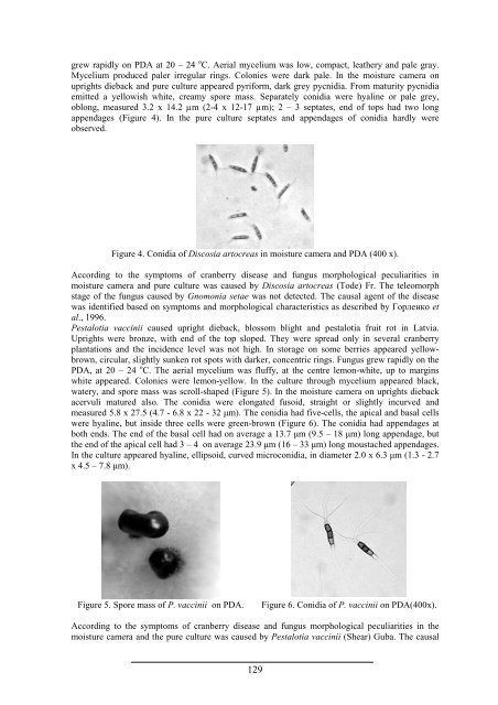
grew rapidly on PDA at 20 – 24 o C. Aerial mycelium was low, compact, leathery and pale gray.Mycelium produced paler irregular rings. Colonies were dark pale. In the moisture camera onuprights dieback and pure culture appeared pyriform, dark grey pycnidia. From maturity pycnidiaemitted a yellowish white, creamy spore mass. Separately conidia were hyaline or pale grey,oblong, measured 3.2 x 14.2 µm (2-4 x 12-17 µm); 2 – 3 septates, end of tops had two longappendages (Figure 4). In the pure culture septates and appendages of conidia hardly wereobserved.Figure 4. Conidia of Discosia artocreas in moisture camera and PDA (400 x).According to the symptoms of cranberry disease and fungus morphological peculiarities inmoisture camera and pure culture was caused by Discosia artocreas (Tode) Fr. The teleomorphstage of the fungus caused by Gnomonia setae was not detected. The causal agent of the diseasewas identified based on symptoms and morphological characteristics as described by Гopлeнкo etal., 1996.Pestalotia vaccinii caused upright dieback, blossom blight and pestalotia fruit rot in Latvia.Uprights were bronze, with end of the top sloped. They were spread only in several cranberryplantations and the incidence level was not high. In storage on some berries appeared yellowbrown,circular, slightly sunken rot spots with darker, concentric rings. Fungus grew rapidly on thePDA, at 20 – 24 o C. The aerial mycelium was fluffy, at the centre lemon-white, up to marginswhite appeared. Colonies were lemon-yellow. In the culture through mycelium appeared black,watery, and spore mass was scroll-shaped (Figure 5). In the moisture camera on uprights diebackacervuli matured also. The conidia were elongated fusoid, straight or slightly incurved andmeasured 5.8 x 27.5 (4.7 - 6.8 x 22 - 32 µm). The conidia had five-cells, the apical and basal cellswere hyaline, but inside three cells were green-brown (Figure 6). The conidia had appendages atboth ends. The end of the basal cell had on average a 13.7 µm (9.5 – 18 µm) long appendage, butthe end of the apical cell had 3 – 4 on average 23.9 µm (16 – 33 µm) long moustached appendages.In the culture appeared hyaline, ellipsoid, curved microconidia, in diameter 2.0 x 6.3 µm (1.3 - 2.7x 4.5 – 7.8 µm).Figure 5. Spore mass of P. vaccinii on PDA.Figure 6. Conidia of P. vaccinii on PDA(400x).According to the symptoms of cranberry disease and fungus morphological peculiarities in themoisture camera and the pure culture was caused by Pestalotia vaccinii (Shear) Guba. The causal129
- Page 3 and 4:
Conference Organizing CommitteeChai
- Page 6 and 7:
15 Pormale J., Osvalde A. and Nolle
- Page 8 and 9:
were established in 1985. Nowadays,
- Page 10 and 11:
10,1-15 ha7%15,1-20 ha7%< 20,1 ha0%
- Page 12 and 13:
In less than half the surveyed farm
- Page 14:
economical and biochemical characte
- Page 17 and 18:
investigated European cranberry acc
- Page 19 and 20:
fruit of V. opulus has different am
- Page 21 and 22:
As several authors have stated (Koz
- Page 23 and 24:
KopsavilkumsVaccinium ăints kultū
- Page 25 and 26:
maintained in a mist chamber with v
- Page 27 and 28:
period and produce vigorous vegetat
- Page 29 and 30:
38. Marcotrigiano M. and McGlew S.P
- Page 31 and 32:
of changes in the typological struc
- Page 33 and 34:
fall from 2 to 3 and that for heath
- Page 35 and 36:
HIGHBUSH BLUEBERRY BREEDINGAUGSTKR
- Page 37 and 38:
Southern and Intermediate highbush
- Page 39 and 40:
and anatomically they belong to fal
- Page 41 and 42:
The levels of flavonols are more co
- Page 43 and 44:
21. Polashock J.J., Griesbach R.J.,
- Page 45 and 46:
Figure 1. A general scheme of the N
- Page 47 and 48:
5. Åkerström A., Forsum Å., Rump
- Page 49 and 50:
species and studying the efficiency
- Page 51 and 52:
Thus, it has been determined that t
- Page 53 and 54:
CHEMICAL COMPOSITION OF HIGHBUSH BL
- Page 55 and 56:
lueberry cultivars were collected f
- Page 57 and 58:
Ascorbic acid, mg 100ḡ 112108642a
- Page 59 and 60:
6. Saftner R., Polashock J., Ehlenf
- Page 61 and 62:
Materials and methodsThe experiment
- Page 63 and 64:
The titrable acids content of the e
- Page 65 and 66:
There was a significant correlation
- Page 67 and 68:
Nichenametla et al., 2006), human n
- Page 69 and 70:
The contribution of V. macrocarpon
- Page 71 and 72:
11. Kong J. M., Chia L. S., Goh N.K
- Page 73 and 74:
isothermically at 70°C for 5 min,
- Page 75 and 76:
IN VITRO PROPAGATION OF SEVERAL VAC
- Page 77 and 78: 16BM ean N o. of shoots/explant1412
- Page 79 and 80: Figure 2. Axillary shoot regenerati
- Page 81 and 82: evaluate the blueberries supply wit
- Page 83 and 84: espectively). It should be stressed
- Page 85 and 86: lueberry appear to play a conclusiv
- Page 87 and 88: 15. Reimann C., Kollen F., Frengsta
- Page 89 and 90: each type, and for comparison sampl
- Page 91 and 92: the mean. Kisgyır 1 sample has the
- Page 93 and 94: 13. Porpáczy A. (1999) A húsos so
- Page 95 and 96: was medium (0.014 - 0.017 g kg -1 s
- Page 97 and 98: ‘Salaspils Ražīgā’. Vigorous
- Page 99 and 100: KopsavilkumsEiropas melleĦu (Vacci
- Page 101 and 102: Figure 2. Chemometric PCA of 32 blu
- Page 103 and 104: References1. Baloga D.W., Vorsa N.,
- Page 105 and 106: obtained from fruits of black choke
- Page 107 and 108: In our opinion, the best estimate a
- Page 109 and 110: cuttings also varies markedly with
- Page 111 and 112: shoots shorter than 10 mm were not
- Page 113 and 114: 14. Ostrolucka M.G., Gajdosova A, L
- Page 115 and 116: „Metos RG-350” (http://www.meto
- Page 117 and 118: 500480Phenols,mg 100g -146044042040
- Page 119 and 120: SHORT INFORMATION ABOUT THE HISTORY
- Page 121 and 122: Evaluation of cultivars. After the
- Page 123 and 124: the number of pistils (female clone
- Page 125 and 126: Table 2. Number of flowers per harv
- Page 127: ResultsFirst time upright dieback i
- Page 131 and 132: Figure 9. Conidia of Physalospora v
- Page 133 and 134: References1. CABI, EPPO, (1997) Dia
- Page 135 and 136: Results und DiscussionBerries were
- Page 137 and 138: In literature Caruso eds. and Гop
- Page 139 and 140: the total area under a cranberry ma
- Page 141 and 142: Skilled works on development of the
- Page 143 and 144: Tika atrastas dažas būtiskas ats
- Page 145 and 146: appears to maintain a quite low lev
- Page 147 and 148: 8. Garkava - Gustavson L.,Persson H