11:10-12:00, Rm 103
11:10-12:00, Rm 103
11:10-12:00, Rm 103
You also want an ePaper? Increase the reach of your titles
YUMPU automatically turns print PDFs into web optimized ePapers that Google loves.
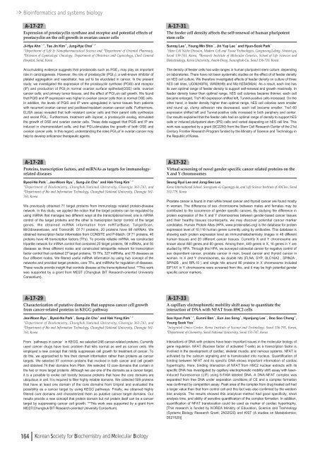
Bioinformatics and systems biologyA-17-27Expression of prostacyclin synthase and receptor and potential effects ofprostacyclin on the cell growth in ovarian cancer cellsJi-Hye Ahn¹ , ², Tae Jin Kim³, Jung-Hye Choi¹ , ²¹Department of Life & Nanopharmaceutical Science and ²Department of Oriental Pharmacy,³Division of Gynecologic Oncology, Department of Obstetrics and Gynecology, Cheil GeneralHospital, Seoul, KoreaAccumulating evidence suggests that prostanoids such as PGE₂may play an importantrole in carcinogenesis. However, the role of prostacyclin (PGI₂), a well-known inhibitor ofplatelet aggregation and vasodilator, has yet to be elucidated in cancer. In the presentstudy, we investigated the expression of the prostacyclin synthase (PGIS) and receptor(IP) and production of PGI₂in normal ovarian surface epithelial(OSE) cells, ovariancancer cells, and primary tumor tissues, and the effect of PGI₂on cell growth. We foundthat PGIS and IP expression was higher in ovarian cancer cells than in normal OSE cells.In addition, the levels of PGIS and IP were upregulated in tumor tissues from patientswith recurrent ovarian cancer and paclitaxel-resistant ovarian cancer cells. Furthermore,ELISA assay revealed that both resistant cancer cells and their parent cells synthesizeand secret PGI₂. Furthermore, treatment with iloprost, a prostacyclin analog, stimulatedthe growth of OSE and ovarian cancer cells. These data suggest that PGIS and IP areinduced in chemoresistant cells, and that PGI₂stimulates the growth of both OSE andovarian cancer cells. In this regard, understanding the roles PGI₂of in ovarian cancer mayhelp to develop anticancer therapeutic agents.A-17-31The feeder cell density affects the self-renewal of human pluripotentstem cellsSunray Lee², Young Min Shin¹, Jin Yup Lee¹and Hyun-Sook Park¹¹Stem Cell Niche Division, Modern Cell and Tissue Technologies, Gongneung2-dong, Nowon-gu,Seoul 139-743, Korea, ²Research Institute of Molecular Genetics, School of Life Sciences andBiotechnology, Korea University, Anam-Dong, Seoungbuk-Gu, Seoul 136-7<strong>10</strong>, KoreaThe density of feeder cells has wide ranges in human pluripotent stem culture, dependingon laboratories. There have not been systematic studies on the effect of of feeder densityon hES cell culture. We therefore investigated effects of feeder density on culture of threehES cell lines, UC06(HSF6), WA09(H9) and Miz-hES4(Miz4). As a result, each line hasits own optimal range of feeder density to support self-renewal and growth maximally. Infeeder density lower than optimal range, hES cell colonies became thinner, each cellbecame enlarged, Tra1-60 expression shifted left, Tunnel-positive cells increased. On theother hand, in feeder density higher than optimal range, hES cell colonies were smallerand round up, clump adhesion rate decreased, each cell became smaller, Tra1-60expression shifted left and Tunnel-positive cells increased in both periphery and center.Our results explained that the feeder cells had an optimal range of density to support hEScells or induced pluripotent stem (IPS) cells and varied depending on hES cell line. Thiswork was supported by a grant (SC2250) from the Stem Cell Research Center of the 21stCentury Frontier Research Program funded by the Ministry of Science and Technology inthe Republic of Korea.A-17-28Proteins, transcription factors, and miRNAs as targets for immunologyrelateddiseasesByeol-Na Park¹, Jea-Woon Ryu¹, Sung-Jin Cho²and Hak-Yong Kim¹ , ²¹Department of Biochemistry, Chungbuk National University, Cheongju 361-763, and²Department of Bio and Information Technology, Chungbuk National University, Cheongju 361-763, KoreaWe previously obtained 71 target proteins from immunology related protein-diseasenetwork. In this study, we applied the notion that the target proteins can be regulated byusing miRNA that manages two different ways at the transcriptional level; one is mRNAcontrol of the target proteins and the other is transcription factor control of the targetgenes. We obtained miRNA information from miRTarBase, TargetScan,Mir2diseasebase, and TransmiR. Of 71 proteins, 20 proteins have 98 miRNAs. Weobtained transcription factor information from CONSITE and P-Match. Of 71 proteins, 46proteins have 48 transcription factors (TFs). To provide reliable miRNA, we constructedtripartite network for mRNA control that contained 20 target proteins, 98 miRNAs, and 56diseases as three different nodes and constructed tetrapartite network for transcriptionfactor control that contained 27 target proteins, 19 TFs, 327 miRNAs, and 78 diseases asfour different nodes. We filtered useful miRNA information by using hub concept of thenetworks and provided target proteins, core TFs, and miRNAs for regulation of diseases.These results provide insight that controls disease at the transcriptional level. **This workwas supported by a grant from MEST (Chungbuk BIT Research-oriented UniversityConsortium).A-17-32Virtual screening of novel gender specific cancer related proteins on theX and Y chromosomesSeung Ryul Lee and Jong-Seo LeeKorea International School, Seongnam-si, Gyeonggi-do, and Life Science Institute of AbClon, Seoul152-779, KoreaProstate cancer is found in man while breast cancer and thyroid cancer are found mostlyin woman. The difference of sex chromosome between males and females may becontributed to the occurrence of gender specific cancers. By studying the difference inprotein expression of the X and Y chromosomes between gender-based cancer tissuesand their healthy tissues counterparts, we may discover potential cancer markercandidates. Human Protein Atlas (HPA, www.proteinatlas.org) is the database for proteinexpression level of <strong>10</strong>,<strong>11</strong>8 human genes currently using by antibodies. This database isshowing each protein expression level as immunohistochemistry images in 48 differenthuman tissues and 20 different cancer tissues. Currently X and Y chromosome areknown about 860 genes and 60 genes. Among them, 449 genes in X, 16 genes in Y arestudied by HPA. Through this HPA, we surveyed colorectal cancer for negative control ofsex dependent cancer, prostate cancer in man, breast cancer and thyroid cancer inwoman. In X and Y chromosomes, six double hits (FLNA, SYP, SLC16A2 , SPIN2A ,SPIN2B , and RPL<strong>10</strong> ) and single hits around 90 proteins in X chromosome includedEIF1AY in Y chromosome were screened from this, and it may be high potential genderspecific cancer markers.A-17-29Characterization of putative domains that suppress cancer cell growthfrom cancer-related proteins in KEGG pathwayJea-Woon Ryu¹, Byeol-Na Park¹, Sung-Jin Cho²and Hak Yong Kim¹ , ²¹Department of Biochemistry, Chungbuk National University, Cheongju 361-763, and²Department of Bio and Information Technology, Chungbuk National University, Cheongju 361-763, KoreaFrom ‘pathways in cancer’in KEGG, we selected 248 cancer-related proteins. Currentlyused cancer drugs have toxic problem that kills normal as well as cancer cells. Weemployed a new concept that mildly suppresses cell growth for treatment of cancer. Todo this, we approached to hire their domain information rather than proteins as cancertargets. We selected 57 common proteins that involved in both cancer and cell growthand obtained 74 their domains from Pfam. We selected <strong>12</strong> core domains that contain inthe two or more target proteins. Although we use one of the domains as a cancer target,it is a possible to evoke cell toxicity because proteins that have the core domains areubiquitous in cell. It is required to filter highly reliable domains. We collected 509 proteinsthat have at least one domain of the core domains from Uniprot and evaluated thepossibility as a cancer target by using KEGG pathways. Finally, we obtained highlyfiltered core domains and characterized them as putative cancer target domains. Ourresults provide a new concept that protein domain but not protein itself can be a cancertarget by suppressing cancer cell growth. **This work was supported by a grant fromMEST(Chungbuk BIT Research-oriented University Consortium).A-17-33A capillary electrophoretic mobility shift assay to quantitate theinteraction of DNA with NFAT from H9C2 cellsSoo Hyun Park¹ , ², Eunmi Ban¹, Eun Joo Song¹, Hyunjung Lee¹, Doo Soo Chung²,Young Sook Yoo¹¹Integrated Omics Center, Korea Institute of Science and Technology, Seoul 136-791, Korea,²Department of Chemistry, Seoul National University, Seoul 151-747, KoreaInteractions of DNA with proteins have been important issues in the molecular biology ofgene regulation. NFAT (Nuclear factor of activated T-cells) as a transcription factor isinvolved in the development of cardiac, skeletal muscle, and nervous systems. NFAT isactivated by the calcium signaling and is translocated into nucleus. Quantification ofbinding between NFAT and its specific DNA shows important information of cardiachypertrophy. Here, binding interaction of NFAT from H9C2 nuclear extracts with itsspecific DNA has investigated by capillary electrophoretic mobility shift assay with laserinducedfluorescence (LIF) using 6-FAM labeled DNA. A DNA-NFAT complex wasseparated from free DNA under separation conditions of CE and a complex formationwas confirmed by competition assay. Peak area of the complex from drug treated cell hada larger value than that from control cell and this fact was also confirmed by the westernblot analysis. The results showed this analytical method had good specificity, shortanalysis time, and ability of sensitive quantification of the complex formation. In addition,quantification of NFAT translocation could be used as marker of cardiac hypertrophy.[This research is funded by KOREA Ministry of Education, Science and Technology(Systems Biology Research Grant; 2N32520) and KIST (A studies on Metabolomics;2E21520)]164 Korean Society for Biochemistry and Molecular Biology




![No 기ê´ëª
(êµë¬¸) ëíì ì íë²í¸ ì¹ì£¼ì ì·¨ê¸í목[ì문] ë¶ì¤ë²í¸ 1 ...](https://img.yumpu.com/32795694/1/190x135/no-eeeeu-e-eii-i-iei-ii-1-4-i-ieiecie-eiei-1-.jpg?quality=85)
