11:10-12:00, Rm 103
11:10-12:00, Rm 103
11:10-12:00, Rm 103
You also want an ePaper? Increase the reach of your titles
YUMPU automatically turns print PDFs into web optimized ePapers that Google loves.
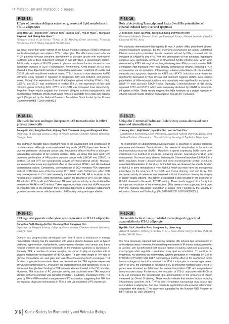
Metabolism and metabolic diseasesP-18-13Effects of Inonotus obliquus extract on glucose and lipid metabolism in3T3-L1 adipocytesJung-Han Lee¹, Eunha Kim¹, Sineun Kim², Sumee Lee², Hye-In Hyun³, TsenguunBayaraa¹and Chang-Kee Hyun¹ , ²¹Graduate School of Life Science, ²School of Life, Handong Global University, ³HandongInternational School, Pohang, Kyungbuk 791-780, KoreaWe have found that water extract of the fungus Inonotus obliquus (IOWE) enhancesinsulin-stimulated glucose uptake in 3T3-L1 adipocytes. This effect was proven to be viaPI-3K pathway through complete abolishment of glucose uptake with wortmannintreatment and a dose dependent increase in Akt activation, a downstream protein.Additionally, analysis of GLUT4 protein in plasma membrane fraction showed a dosedependent increase in GLUT4 translocation. Furthermore, IOWE-treated 3T3-L1 cellsshowed a dose dependent increase in adiponectin expression. Treatment of HepG2 andC2C<strong>12</strong> cells with conditioned media of treated 3T3-L1 induced a dose dependent AMPKactivation, a key regulator in regulation of lipogenesis, fatty acid oxidation, and glucoseintake. Though the expression of several adipogenic genes including PPARγ, FAS,C/EBPα, and SCD1 was unaffected in treated 3T3-L1, the expression of fatty acidoxidation genes including AOX, CPT1, and LCAD was increased dose dependently.Together, these results suggest that Inonotus obliquus exhibits hypoglycemic andbeneficial lipid metabolic effects which would make it a candidate for a viable anti-diabeticagent. [Supported by the National Research Foundation Grant funded by the KoreanGovernment (MEST, 2<strong>00</strong>9-<strong>00</strong>65952)].P-18-16Role of Activating Transcriptional Factor 3 on HBx potentiation ofethanol-induced fatty liver and apoptosisJi Yeon Kim, Keon Jae Park, Jeong Suk Kang and Won-Ho KimDivision of Metabolic Diseases, Center for Biomedical Science, National Institute of Health,Chungbuk 363-951, KoreaWe previously demonstrated that hepatitis B virus X protein (HBx) potentiated ethanolinducedhepatocyte apoptosis, but the underlying mechanisms are poorly understood.Ethanol consumption potentiated hepatic steatosis induced by HBx, correlated with theinduction of SREBP1c and FAS. With the potentiation of liver steatosis, hepatocyteapoptosis was significantly increased in ethanol-fed AdHBx-infected mice, which weredetermined by ATF3. Although ethanol negatively regulated IFN-γproduction unlike TNFαinduction,HBx-mediated IFN-γwas potently enhanced by ethanol, following STAT1accumulation via its activation. Interestingly, ethanol potentiation of HBx-mediatedsteatosis and apoptosis depends on ATF3 and STAT1 induction since these aresignificantly decreased by their siRNAs and dominant negative cDNAs. Also, ethanolpotentiation of HBx-induced steatosis and apoptosis was significantly increased inSTAT1+/+ mice, but not in STAT1-/- mice. Especially, C-terminal domain of HBx directlyregulated ATF3 and STAT1, which were completely abolished by HBxNT or tetracyclinoffsystem of HBx. These results suggest that HBx functions as a potent regulator ofethanol-induced hepatic steatosis and apoptosis through ATF3 induction.P-18-14Oleic acid induces androgen-independent AR transactivation in 22Rv1prostate cancer cellsSeung-Jin Kim, Sung-Soo Park, Hojung Choi, Yoonseok Jung and Eungseok KimDepartment of Biological Sciences, College of Natural Sciences, Chonnam National University,Gwangju, KoreaThe androgen receptor plays important roles in the development and progression ofprostate cancer. Although monounsaturated fatty acids (MUFA) have been known topromote proliferation of prostate cancer cells, the potential effect of MUFAs on androgenindependentactivation of AR remains unclear. Here, we show that oleic acid (OA)promotes proliferation of AR-positive prostate cancer cells (LNCaP and 22Rv1). Inaddition, OA and DHT can synergistically activate AR transcriptional activity. However,we were not able to see any significant effect of oleic acid on PPARγ- and GR-mediatedtranscriptional activity. Consistently, overexpression of SCD increases PSA expressionand cell proliferation even at the low level of DHT (0.01~1 nM). Furthermore, when SCDwas overexpressed in CV1 cells transiently transfected with AR, AR is localized in thenucleus at 0.01 nM DHT. More interestingly, even in the absence of DHT, OA can induceAR activity in 22Rv1 cells and OA-induced AR transcriptional activity can be abolished bytreatment of MAPK or AKT inhibitor. Taken together, our data show that MUFA may playan important role in the transition from androgen-dependent to androgen-independentgrowth of prostate cancer cells by activation of AR activity during androgen ablation.P-18-17Ubiquitin C-terminal Hydrolase-L3 deficiency causes decreased bonemass and mineralizationJi Young Kim¹, Keiji Wada², Hye-Sim Cho¹and Je-Yoel Cho¹¹Department of Biochemistry, School of Dentistry, Kyungpook National University, Daegu, Korea,²National Institute of Neuroscience, National Center of Neurology & Psychiatry, Tokyo, JapanThe mechanism of ubiquitination/deubiquitination is essential in various biologicalprocesses and diseases. Deubiquitination, the reversal of ubiquitination, is the action ofdeubiquitinating enzymes (DUBs). Mutations in genes expressing DUBs have beenimplicated in a number of diseases including cancer, neurodegeneration, andosteoporosis. Our recent study showed that ubiquitin C-terminal hydrolase L3 (Uch-L3), aDUB, regulates Smad1 ubiquitination and bone morphogenetic protein 2-inducedosteoblast differentiation. In this study, for the first time, we observed the specific functionof Uch-L3 in bone metabolism in vivo. Uch-L3 knock-out mice have the osteoporoticphenotype by the analysis of micro-CT, von Kossa staining, and soft X-ray. Thedecreased activity of osteoblast was observed in Uch-L3 knock-out mice by the analysisof calcein double labeling. The activity of osteoclast is also elevated in the deficiency ofUch-L3, observed in the result of TRAP staining. These results suggest Uch-L3 might bean important modulator of bone metabolism. [This research was supported by a grantfrom the National Research Foundation of Korea (NRF) funded by the Ministry ofeducation, Science and Technology (No. 20<strong>10</strong>-<strong>00</strong><strong>11</strong>231 & 20<strong>10</strong>-<strong>00</strong>20553).]P-18-15TR4 regulates pyuvate carboxylase gene expression in 3T3-L1 adipocytesSung-Soo ParK, Seung-Jin Kim, Ho-Jung Choi, Eungseok KimDepartment of Biological Sciences, College of Natural Sciences, Chonnam National University,Gwangju, KoreaObesity has progressively developed over time if there is imbalance in energyhomeostasis. Obesity has the association with various chronic diseases such as type 2diabetes, hypertension, dyslipidemia, cardiovascular disease, and cancer and thesemedical problems are collectively referred to as the metabolic syndrome or Syndrome X.Recently, TR4, a member of nuclear receptor, has shown to play an important role inglucose metabolism via regulation of PEPCK gene. To gain more insight of TR4 role inglucose homeostasis, we used gain- and loss-of-function approaches to investigate TR4function on glucose homeostasis. Here, we demonstrate that TR4 regulates expressionof Pyruvate carboxylase(PC), involved in the gluconeogenesis and lipogenesis, in 3T3-L1adipocytes through direct binding to TR4 response element located in the PC promoter.Moreover, TR4 induction of PC promoter activity was abolished when TR4 responseelement in the PC promoter was disrupted (mutated). In addition, knockdown of the TR4gene by TR4 miRNA resulted in suppression of PC expression, suggesting that TR4 is akey regulator of glucose homeostasis in 3T3-L1 cells via modulation of PC expression.P-18-18The soluble factors from γ-irradiated macrophages trigger lipidaccumulation in 3T3-L1 adipocyteHye Min Choi*, Hae-Ran Park, Sung-Kee Jo, Uhee-JungAdvanced Radiation Technology Institute (ARTI), Korea Atomic Energy Institute (KAERI),Jeonbuk 580-185, KoreaWe have previously reported that ionizing radiation (IR) induces lipid accumulation inwhite adipose tissue. However, the underlying mechanism of IR-induce lipid accumulationis unclear. We hypothesized that soluble factors including cytokines produced bymacrophges after regulate γ-irradiation may lipid accumulation. To confirm ourhypothesis, we examined the inflammatory cytokine production in γ-irradiated (IR-CM) orLPS-trated (LPS-CM) RAW 264.7 macrophages and the effect of the conditioned mediaby macrophages on the lipid accumulation in 3T3-L1 adipocytes. In macrophages treatedwith IR or LPS, the expression of interleukin-6 (IL-6) and tumor necrosis factor α(TNF-α)genes were increased as determined by reverse transcription PCR and enzyme-linkedimmunosorbent assay. Furthermore, the incubation of 3T3-L1 adipocytes with IR-CM orLPS-CM increased the intracellular lipid accumulation in the presence of insulinmeasured by Oil-red O staining. These results indicate that soluble factors includinginflammatory cytokines (IL-6, TNF-α) from γ-irradiated macrophage may induce lipidaccumulation in adipocytes. And thus contribute significantly to the systemic inflammationassociated with obesity. [This study was supported by the Nuclear R&D Program ofMEST (Grant No. 2<strong>00</strong>7-2<strong>00</strong><strong>00</strong>91)]316 Korean Society for Biochemistry and Molecular Biology



![No 기ê´ëª
(êµë¬¸) ëíì ì íë²í¸ ì¹ì£¼ì ì·¨ê¸í목[ì문] ë¶ì¤ë²í¸ 1 ...](https://img.yumpu.com/32795694/1/190x135/no-eeeeu-e-eii-i-iei-ii-1-4-i-ieiecie-eiei-1-.jpg?quality=85)
