11:10-12:00, Rm 103
11:10-12:00, Rm 103
11:10-12:00, Rm 103
Create successful ePaper yourself
Turn your PDF publications into a flip-book with our unique Google optimized e-Paper software.
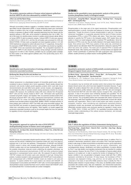
ProteomicsS-18-01The protein expression patterns of human retinal pigment epitheliumderived ARPE-19 cells were attenuated by oxidative stressJi-Eun Lee and Kee Ryeon KangDepartment of Biochemistry and MRC for Neural Dysfunction, School of Medicine and Institute ofHealth Sciences, Gyeongsang National University, Jinju 660-751, KoreaAlthough accumulating evidence suggests a correlation between oxidative stress andage-related macular degeneration (AMD), a causal relationship based upon mechanisticstudies of oxidative injury has not been established. Identification of proteins whosefunction or expression is altered in AMD, especially determining how they interact with theapoptotic pathway in RPE cells, will be essential in establishing their role in AMD. Theaim of this study is to identify proteins involved in oxidative stress-induced cellular effectson human ARPE-19 cells by proteomic analysis. Cultured ARPE-19 cells were exposedto H2O2 (0.5 to 2 mM) for 24 h, and then the cell lysates were processed for 2-DE andMALDI-TOF/MS. Of these spots up-regulated and down-regulated proteins in 0.5 mMH2O2 treatment-as compared to control cells-were 42 and 17, respectively. Among themthe expression of MAP RP/EB family member 1 and prohibitin was showed up-regulated,and HnRNP C1/C2 was expressed down-regulated. The up-regulated expression ofprohibitin on ARPE-19 cells showed consistency during hydrogen peroxide treatmentranging from 0.5 mM to 2 mM. Therefore, prohibitin may be a possible protein candidatefor ROS marker in the retinal pigment epithelial cells of the eye.[Funding source: R13-2<strong>00</strong>5-0<strong>12</strong>-0<strong>10</strong>03-0]S-18-04Studies on the quantitative mass spectrometric analysis of the proteintyrosine phosphatase family in biological sampleTae-Jin jeon¹, Jeong-Hee Moon², Dae-gwin Jeong², Tae-Sung Yoon², Young-JaeBahn²and Seong-Eon Ryu¹¹Department of Life Science, Hanyang University, Seoul 133-791, Korea, ²Medical ProteomicsResearch Center, KRIBB, Daejeon 305-806, KoreaPhosphorylation is the major part of the signal transduction. Phosphorylation anddephosphorylation is strictly controlled and balanced by kinase and phosphatase,respectively. Though the amount of tyrosin phosphorylation is quite low, it has beenintensively investigated. It is generally assumed that the level of Protein tyrosinephosphatases (PTPs) or active PTPs reflects cells status. There have been variousattempts to quantify the PTP levels in the biological system. EGF treated HeLa cellsystem is chosen as model biological system to optimize PTP quantification. Since theabundance of PTPs is very low, they are hardly detected in the trypsin treated whole celllysate. Since almost every PTPs have cysteine at their active site, enrichment bycysteine-capture was attempted. Biotin-PEO-iodoacetamide is utilized for capturing PTPswhich has active site cysteine. The biotin part of captured PTPs is reacted withStreptavidin beads. After washing and centrifugation, enriched proteins are digested withtrypsin. Protein quantification is performed with triple quadrupole mass spectrometer(Waters, Quattro premiere) by multiple reaction monitoring(MRM) method.S-18-02Identification and characterization of ionizing radiation-inducedsenescence associated secretomeNa-Kyung Han, Bong-Cho Kim and Jae-Seon LeeDivision of Radiation Cancer Research, Korea Institute of Radiological and Medical Sciences, Seoul139-706, KoreaCellular senescence is a physiological program of irreversible growth arrest, which isconsidered to play an important role in tumor suppression. Recent studies showed thatsenescent cells secrete multiple growth-regulatory proteins which could alter tissuemicroenvironments and could affect tumor growth, survival, invasion, and angiogenesis.To investigate the effect of secreted proteins during senescence induction by ionizingradiation (IR) exposure, the conditioned media (CM) were collected from IR-exposedMCF7 cells and then directly treated to MCF7 cells. CM-treated cells significantlyincreased cell growth, invasion, and migration. Next, we examined secreted proteins fromIR-induced senescent MCF7 cells using 2-dimensional electrophoresis combined withMALDI-TOF MS. As a result, we identified 34 differentially secreted protein spots andselected novel secreted proteins including RKIP, MARK4, AKAP9. Increased secretion ofthose proteins were confirmed in MCF7 and H460 cells using immunoblot analysis. Wefound that RKIP was secreted via classical secretion pathway and secreted RKIP playscritical role in stimulating cancer cell migration. Taken together, our results suggest thatsenescence associated secretome could be the principal targets that have the potentialfor regulation of tumor cell growth and migration.S-18-05Quantitative proteomic analysis of differentially secreted proteins indesigned isogenic breast cancer cell linesUn-Beom Kang¹, Hyeong-Gon Moon², Eunmi Ban¹, Jin-Young Kim¹, YoonKyoung Jun¹, Dong-Young Noh², Hye-Seoung Shin¹¹Bio-Medieng Inc., 333-7 Sangdaewon 1-dong, Jungwon-gu, Seongnam-si, Gyeonggi-do, Korea,²Cancer Research Institute, Seoul National University College of Medicine, Seoul, KoreaThe secretome may be enriched with secreted and/or shed proteins from adjacentdisease-relevant cancer cells, which have been targeted for biomarker discovery. To gaininsights into proteins released from cells and obtain breast cancer related proteins, wecompared secretome profiles between nontumorigenic MCF<strong>10</strong>A cells and tumorigenicRas-transformed MCF<strong>10</strong>A cells that stably express oncogenic Ras, using SILACapproach. A total of 343 proteins were confidently identified and quantified. The 46proteins showed abundance changes by more than 1.5-fold between the isogenic breastcancer cell lines. The up-regulated proteins were associated with cellular movement andcell morphology, while down-regulated proteins were predominantly involved with cellularassembly and organization. About a half of total protein was mainly occupied oncytoplasm, however, when considering only significantly expressed proteins,cyproplasmic protein was decreased, and extracellular space was raised. Several ofdifferentially secreted proteins were previously reported as regulated in breast cancer(e.g. TGFB2, CXCL1, S<strong>10</strong>0A2, and GSTP1). Our quantitative secretome profiles mayprovide a better understanding of the cellular process mechanism, and clinicallymeaningful target proteins.S-18-03The proteomic approach to explore the roles of HAUSP/USP7deubiquitinating enzyme in human cancer and apoptosisKey-Hwan Lim, Jun-Hyun Kim, Suresh Ramakrishna, Ji-Hyun Yun, Ji-Hee Kim andKwang-Hyun BaekDepartment of Biomedical Science, CHA University, CHA Stem Cell Institute, Seoul 135-081,KoreaHAUSP (Herpes-virus-associated ubiquitin-specific protease, USP7) is a deubiquitinatingenzyme and has activity in stabilizing tumor suppressor protein p53, and in turn plays animportant role in cell cycle and apoptosis. Overexpression of hHAUSP and hHAUSP(C224S) mutant in cancer cells revealed apoptotic phenotype, although hHAUSP(C224S) mutation does not have deubiquitinating enzyme activity. Therefore, to explorethe possible intracellular molecular events underlining the apoptotic phenotype, weperformed two-dimensional electrophoresis (2-DE) and other proteomics-basedapproaches in overexpressed human cancer cells. We analyzed 35 spots by MALDI-TOF/TOF analysis and 26 protein spots were detected in each gel. Among theseproteins, 4 proteins revealed in cells overexpressed with hHUASP and 3 novel proteins incells overexpressed with a hHAUSP (C224S) mutant are related to cell cycle andapoptosis. To explore whether the 4 novel proteins can interact with hHAUSP, weoverexpressed 4 novel proteins and hHAUSP in 293T cells. Co-immunoprecipitations anddeubiquitination assays revealed that these novel 4 proteins interact with hHAUSP, whichcan deubiquitinate them. Functional studies are unclear investigation.S-18-06Prdx V affects the regulation of kidney homeostasis during hypoxiaHee-Young Yang¹, Joseph Kwon², Hoon-In Choi¹, Lina Ren¹, Ung Yang¹, Byung-Ju Park and Tae-Hoon Lee¹¹Department of Oral Biochemistry, Dental Science Research Institute, The 2nd Stage of BrainKorea 21 for Dental School, Chonnam National University, Gwangju 5<strong>00</strong>-757, Korea, ²Korea BasicScience Institute, Gwangju 5<strong>00</strong>-757, KoreaPeroxiredoxin V (Prdx V) is widely expressed in mammalian tissues. In addition, Prdx V islocalized in mitochondria, peroxisome, cytosol, and nucleus. Prdx V has been reported toprotect a wide range of cellular environments as antioxidant enzyme, and its dysfunctionsmay be implicated in several diseases, such as cancer, inflammation, andneurodegenerative disease. Identification and relative quantification of proteins affectedby Prdx V may help identify novel signaling mechanisms that are important for oxidativestress response. However, the role of Prdx V in the modulation of hypoxia-related cellularresponse is not studied yet. In order to examine the function of endogenous Prdx V inhypoxic condition in vivo, we generated a transgenic mouse model with Prdx V siRNAexpression controlled by U6 promoter. Of many tissues, the knockdown of Prdx Vexpression was displayed in kidney, lung, and liver, but not spleen and skin. Weconducted on the basis of nano-UPLC-MSE proteomic study to identify the Prdx V-affected protein networks in hypoxic kidneys. In this study, we identified protein networksassociated with oxidative stress, fatty acid metabolism, and mitochondrial dysfunction.Our results indicated that Prdx V affected to regulation of kidney homeostasis underhypoxia stress.332 Korean Society for Biochemistry and Molecular Biology




![No 기ê´ëª
(êµë¬¸) ëíì ì íë²í¸ ì¹ì£¼ì ì·¨ê¸í목[ì문] ë¶ì¤ë²í¸ 1 ...](https://img.yumpu.com/32795694/1/190x135/no-eeeeu-e-eii-i-iei-ii-1-4-i-ieiecie-eiei-1-.jpg?quality=85)


