11:10-12:00, Rm 103
11:10-12:00, Rm 103
11:10-12:00, Rm 103
You also want an ePaper? Increase the reach of your titles
YUMPU automatically turns print PDFs into web optimized ePapers that Google loves.
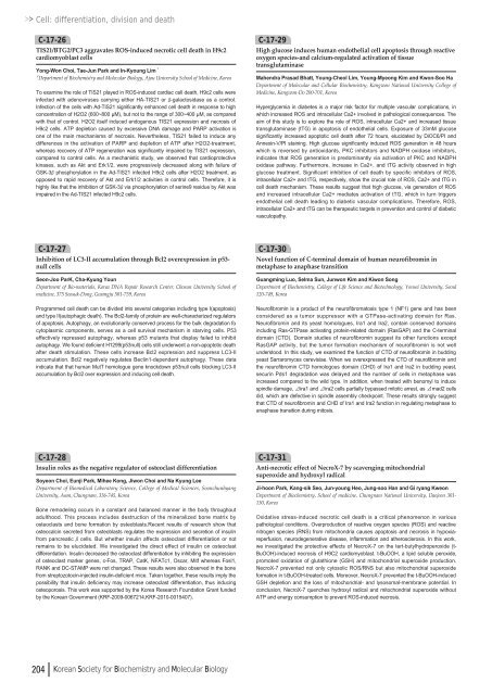
Cell: differentiation, division and deathC-17-26TIS21/BTG2/PC3 aggravates ROS-induced necrotic cell death in H9c2cardiomyoblast cellsYong-Won Choi, Tae-Jun Park and In-Kyoung Lim¹¹Department of Biochemistry and Molecular Biology, Ajou University School of Medicine, KoreaTo examine the role of TIS21 played in ROS-induced cardiac cell death, H9c2 cells wereinfected with adenoviruses carrying either HA-TIS21 or β-galactosidase as a control.Infection of the cells with Ad-TIS21 significantly enhanced cell death in response to highconcentration of H2O2 (6<strong>00</strong>~8<strong>00</strong> µM), but not to the range of 3<strong>00</strong>~4<strong>00</strong> µM, as comparedwith that of control. H2O2 itself induced endogenous TIS21 expression and necrosis ofH9c2 cells. ATP depletion caused by excessive DNA damage and PARP activation isone of the main mechanisms of necrosis. Nevertheless, TIS21 failed to induce anydifferences in the activation of PARP and depletion of ATP after H2O2-treatment,whereas recovery of ATP regeneration was significantly impaired by TIS21 expression,compared to control cells. As a mechanistic study, we observed that cardioprotectivekinases, such as Akt and Erk1/2, were progressively decreased along with failure ofGSK-3βphosphorylation in the Ad-TIS21 infected H9c2 cells after H2O2 treatment, asopposed to rapid recovery of Akt and Erk1/2 activities in control cells. Therefore, it ishighly like that the inhibition of GSK-3βvia phosphorylation of serine9 residue by Akt wasimpaired in the Ad-TIS21 infected H9c2 cells.C-17-29High glucose induces human endothelial cell apoptosis through reactiveoxygen species-and calcium-regulated activation of tissuetransglutaminaseMahendra Prasad Bhatt, Young-Cheol Lim, Young-Myeong Kim and Kwon-Soo HaDepartment of Molecular and Cellular Biochemistry, Kangwon National University College ofMedicine, Kangwon-Do 2<strong>00</strong>-701, KoreaHyperglycemia in diabetes is a major risk factor for multiple vascular complications, inwhich increased ROS and intracellular Ca2+ involved in pathological consequences. Theaim of this study is to explore the role of ROS, intracellular Ca2+ and increased tissuetransglutaminase (tTG) in apoptosis of endothelial cells. Exposure of 33mM glucosesignificantly increased apoptotic cell death after 72 hours, elucidated by DiOC6/PI andAnnexin-V/PI staining. High glucose significantly induced ROS generation in 48 hourswhich is reversed by antioxidants, PKC inhibitors and NADPH oxidase inhibitors,indicates that ROS generation is predominantly via activation of PKC and NADPHoxidase pathway. Furthermore, increase in Ca2+, and tTG activity observed in highglucose treatment. Significant inhibition of cell death by specific inhibitors of ROS,intracellular Ca2+ and tTG, respectively, show the crucial role of ROS, Ca2+ and tTG incell death mechanism. These results suggest that high glucose, via generation of ROSand increased intracellular Ca2+ mediates activation of tTG, which in turn triggersendothelial cell death leading to diabetic vascular complications. Therefore, ROS,intracellular Ca2+ and tTG can be therapeutic targets in prevention and control of diabeticvasculopathy.C-17-27Inhibition of LC3-II accumulation through Bcl2 overexpression in p53-null cellsSeon-Joo ParK, Cha-Kyung YounDepartment of Bio-materials, Korea DNA Repair Research Center, Chosun University School ofmedicine, 375 Seosuk-Dong, Gwangju 501-759, KoreaProgrammed cell death can be divided into several categories including type I(apoptosis)and type II(autophagic death). The Bcl2-family of protein are well-characterized regulatorsof apoptosis. Autophagy, an evolutionarily conserved process for the bulk degradation focytoplasmic components, serves as a cell survival mechanism in starving cells. P53effectively repressed autophagy, whereas p53 mutants that display failed to inhibitautophagy. We found deficient H<strong>12</strong>99(p53null) cells still underwent a non-apoptotic deathafter death stimulation. These cells increase Bcl2 expression and suppress LC3-IIaccumulation. Bcl2 negatively regulates Beclin1-dependent autophagy. These dataindicate that that human MutT homologue gene knockdown p53null cells blocking LC3-IIaccumulation by Bcl2 over expression and inducing cell death.C-17-30Novel function of C-terminal domain of human neurofibromin inmetaphase to anaphase transitionGuangming Luo, Selma Sun, Junwon Kim and Kiwon SongDepartment of Biochemistry, College of Life Science and Biotechnology, Yonsei University, Seoul<strong>12</strong>0-749, KoreaNeurofibromin is a product of the neurofibromatosis type 1 (NF1) gene and has beenconsidered as a tumor suppressor with a GTPase-activating domain for Ras.Neurofibromin and its yeast homologues, Ira1 and Ira2, contain conserved domainsincluding Ras-GTPase activating protein-related domain (RasGAP) and the C-terminaldomain (CTD). Domain studies of neurofibromin suggest its other functions exceptRasGAP activity, but the tumor formation mechanism of neurofibromin is not wellunderstood. In this study, we examined the function of CTD of neurofibromin in buddingyeast Sarraromyces cerevisiae. When we overexpressed the CTD of neurofibromin andthe neurofibromin CTD homologous domain (CHD) of Ira1 and Ira2 in budding yeast,securin Pds1 degradation was delayed and the number of cells in metaphase wasincreased compared to the wild type. In addition, when treated with benomyl to inducespindle damage, Δira1 and Δira2 cells partially bypassed mitotic arrest, as Δmad2 cellsdid, which are defective in spindle assembly checkpoint. These results strongly suggestthat CTD of neurofibromin and CHD of Ira1 and Ira2 function in regulating metaphase toanaphase transition during mitosis.C-17-28Insulin roles as the negative regulator of osteoclast differentiationSoyeon Choi, Eunji Park, Mihee Kong, Jiwon Choi and Na Kyung LeeDepartment of Biomedical Laboratory Science, College of Medical Sciences, SoonchunhyangUniversity, Asan, Chungnam, 336-745, KoreaBone remodeling occurs in a constant and balanced manner in the body throughoutadulthood. This process includes destruction of the mineralized bone matrix byosteoclasts and bone formation by osteoblasts.Recent results of research show thatosteocalcin secreted from osteoblasts regulates the expression and secretion of insulinfrom pancreatic βcells. But whether insulin affects osteoclast differentiation or notremains to be elucidated. We investigated the direct effect of insulin on osteoclastdifferentiation. Insulin decreased the osteoclast differentiation by inhibiting the expressionof osteoclast marker genes, c-Fos, TRAP, CatK, NFATc1, Oscar, Mitf whereas Fosl1,RANK and DC-STAMP were not changed. These results were also observed in the bonefrom streptozotocin-injected insulin-deficient mice. Taken together, these results imply thepossibility that insulin deficiency may increase osteoclast differentiation, thus inducingosteoporosis. This work was supported by the Korea Research Foundation Grant fundedby the Korean Government (KRF-2<strong>00</strong>9-<strong>00</strong>67214,KRF-20<strong>10</strong>-<strong>00</strong>15407).C-17-31Anti-necrotic effect of NecroX-7 by scavenging mitochondrialsuperoxide and hydroxyl radicalJi-hoon Park, Kang-sik Seo, Jun-young Heo, Jung-soo Han and Gi ryang KweonDepartment of Biochemistry, School of medicine, Chungnam National University, Daejeon 301-130, KoreaOxidative stress-induced necrotic cell death is a critical phenomenon in variouspathological conditions. Overproduction of reactive oxygen species (ROS) and reactivenitrogen species (RNS) from mitochondria causes apoptosis and necrosis in hypoxiareperfusion,neurodegenerative disease, inflammation and atherosclerosis. In this work,we investigated the protective effects of NecroX-7 on the tert-butylhydroperoxide (t-BuOOH)-induced necrosis of H9C2 cardiomyoblast. t-BuOOH, a lipid soluble peroxide,promoted oxidation of glutathione (GSH) and mitochondrial superoxide production.NecroX-7 prevented not only cytosolic ROS/RNS but also mitochondrial superoxideformation in t-BuOOH-treated cells. Moreover, NecroX-7 prevented the t-BuOOH-inducedGSH depletion and the loss of mitochondrial- and lysosomal-membrane potential. Inconclusion, NecroX-7 quenches hydroxyl radical and mitochondrial superoxide withoutATP and energy consumption to prevent ROS-induced necrosis.204 Korean Society for Biochemistry and Molecular Biology


![No 기ê´ëª
(êµë¬¸) ëíì ì íë²í¸ ì¹ì£¼ì ì·¨ê¸í목[ì문] ë¶ì¤ë²í¸ 1 ...](https://img.yumpu.com/32795694/1/190x135/no-eeeeu-e-eii-i-iei-ii-1-4-i-ieiecie-eiei-1-.jpg?quality=85)
