11:10-12:00, Rm 103
11:10-12:00, Rm 103
11:10-12:00, Rm 103
Create successful ePaper yourself
Turn your PDF publications into a flip-book with our unique Google optimized e-Paper software.
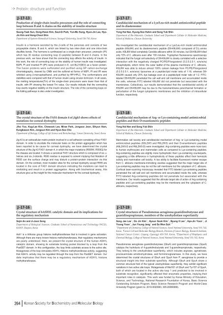
Protein: structure and functionJ-17-13Production of single-chain insulin precursors and the role of connectingloop between B and A chains on the stability of insulin structureSeung-Taek Sun, Dong-Hwan Kim, Seul-Gi Park, Yun-Mo Sung, Hyun-Jin Lee, Hyo-Jin Kim and Hang-Cheol ShinDepartment of Systems Biomedical Sciences, Soongsil University, Seoul 156-743, KoreaInsulin is a hormone secreted by the β-cells of the pancreas and consists of twopolypeptide chains, B and A, which are linked by two inter-chain and one intra-chaindisulfide bonds. The hormone is synthesized as a single-chain precursor, proinsulin (PI)which has 35 residue connecting loop (C-peptide) between B and A chains. In theprevious work, we have shown that the connecting loop affects the activity of insulin. Inthis work, the role of connecting loop on the stability of human insulin was investigated.M2PI, PI and inverted PI (IPI) were produced in E. coli BL21(DE3) as a fusion protein.The fusion proteins were sulfonated and purified using SP cation-exchangechromatography, cleaved by CNBr and the sulfonat ed forms of M2PI, PI and IPI wererefolded using β-mercaptoethanol, and purified by RP-HPLC. The conformations andstabilities were compared with that of human insulin using circular dichroism. In all cases,the melting temperatures(Tm) of the single-chain precursors were lower than that ofinsulin, with IPI showing the lowest Tm value. Our results indicate that the connectingloop exerts negative stability on the insulin structure. The role of the connecting loops onthe folding pathways is also under investigation.J-17-17Candidacidal mechanism of a Lys/Leu-rich model antimicrobial peptideand its diastereomerYong Hai Nan, Kyung-Soo Hahm and Song Yub ShinDepartment of Bio-Materials, Graduate School and Department Cellular & Molecular Medicine,School of Medicine, Chosun UniversityWe investigated the candidacidal mechanism of a Lys/Leu-rich model antimicrobialpeptide (K9L8W) and its diastereomeric peptide (D9-K9L8W) composed of D,L-aminoacids. K9L8W killed completely Candida albicans within 30 minutes, but D9-K9L8W killedonly 72% of C. albicans even after <strong>10</strong>0 minutes. Tryptophan fluorescence spectroscopyindicated that the fungal cell selectivity of D9-K9L8W is closely correlated with a selectiveinteraction with the negatively charged PC/PE/PI/ergosterol (5:2.5:2.5:1, w/w/w/w)phospholipids, which mimic the outer leaflet of the plasma membrane of C. albicans.K9L8W was able to induce almost <strong>10</strong>0% calcein leakage from PC/PE/PI/ergosterol(5:2.5:2.5:1, w/w/w/w) liposomes at a peptide:lipid molar ratio of 1:16, whereas D9-K9L8W caused only 25% dye leakage even at a peptide:lipid molar ratio of 1:2. FITClabeledD9-K9L8W penetrated the cell wall and cell membrane and accumulated insidethe cells, whereas FITC-labeled K9L8W did not penetrate but associated with themembranes. Collectively, our results demonstrated that the candidacidal activity ofK9L8W and D9-K9L8W may be due to the transmembrane pore/channel formation orperturbation of the fungal cytoplasmic membranes and the inhibition of intracellularfunction, respectively.J-17-14The crystal structure of the FAS1 domain 4 of βigh3 shows effects ofmutations for corneal dystrophyJiho Yoo, Kug-Lae Kim, Chewook Lee, Mirae Park, Jongsoo Jeon, Sihyun Ham,Eungkweon Kim, Jongsun Kim and Hyun-Soo ChoDepartment of Biology, College of Life Science and Biotechnology, Yonsei University, Seoul, Koreaβig-h3 is an extracellular matrix protein involved in a cell adhesion consisting of four FAS1domain. In order to elucidate the molecular basis on the protein aggregation which hasbeen reported to be cause for corneal dystrophy, we have determined the crystalstructure of the βig-h3 FAS1 domain 4, in which the major mutations (R555W, R555Q) forthe disease are located. It shows a canonical FAS1 structure which is composed of six α-helices and six β-strands. R555 residue is exposed to solvent implying that Mutation ofR555 can the surface charge and may disturb a protein-protein interaction via thisdomain. On the contrast, most mutation sites for the corneal dystrophy except R555 arelocated in the core of FAS1 domain structure indicating the mutations can lead tomisfolding and result in a protein aggregation. Along with biochemical assay, thisstructure give us the insight for the molecular mechanism for the corneal dystrophy.J-17-18Candidacidal mechanism of Arg- or Lys-containing model antimicrobialpeptides and their D-enantiomeric peptidesYong Hai Nan and Song Yub ShinDepartment of Bio-Materials, Graduate School and Department Cellular & Molecular Medicine,School of Medicine, Chosun UniversityMammalian cell toxicity and candidacidal mechanism of Arg- or Lys-containing modelantimicrobial peptides (K6L2W3 and R6L2W3) and their D-enantiomeric peptides(K6L2W3-D and R6L2W3-D) were investigated. Arg-containing peptides were more toxicto human erythrocytes and mammalian cells as compared to Lys-containing peptides.Arg-containing peptides are slightly more hydrophobic than Lys-containing counterparts,suggesting a little difference in hydrophobicity of these peptides affect their hemolyticactivity and mammalian cell toxicity. A low ability to facilitate fluorescent marker escapefrom C. albicans membrane-mimicking vesicles suggested that the major target site ofLys-containing peptides may be not the cell membrane but the cytoplasm of C. albicans.Confocal laser-scanning microscopy revealed that FITC-labeled Lys-containing peptidespenetrated the cell wall and cell membrane and accumulated inside the cells, whereasFITC-labeled Arg-containing peptides did not penetrate but associated with themembrane. Our results suggested that the ultimate target site of action of Arg-containingpeptides and Lys-containing peptides may be the membrane and the cytoplasm of C.albicans, respectively.J-17-16Crystal structure of hASH1L catalytic domain and its implications forthe regulatory mechanismSojin An and Ji-Joon SongDepartment of Biological Sciences, Graduate School of Nanoscience and Technology (WCU),KAIST, Daejeon, KoreaAsh1 is a trithorax group histone methyltransferase that is involved in gene activation.Although there are many known histone methyltransferases, their regulatory mechanismsare poorly understood. Here, we present the crystal structure of the human ASH1Lcatalytic domain, showing its substrate binding pocket blocked by a loop from thePostSET domain. In this configuration, the loop limits substrate access to the active site.Mutagenesis of the loop stimulates ASH1L Histone methyltransferase activity, suggestingthat ASH1L activity may be regulated through the loop from the PostSET domain. Ourdata implicates that there may be a regulatory mechanism of ASH1L histonemethyltransferases.J-17-19Crystal structures of Pseudomonas aeruginosa guanidinobutyrase andguanidinopropionase, members of the ureohydrolase superfamilySang Jae Lee¹, Do Jin Kim¹, Hyoun Sook Kim¹, Byung Il Lee², Hye-Jin Yoon¹, JiYoung Yoon¹, Jun Young Jang¹and Se Won Suh¹ , ³¹Department of Chemistry, College of Natural Sciences, Seoul National University, Seoul 151-742,Korea, ²Cancer Cell and Molecular Biology Branch, Division of Cancer Biology, Research Institute,National Cancer Center, Goyang, Gyeonggi 4<strong>10</strong>-769, Korea, ³Department of Biophysics andChemical Biology, College of Natural Sciences, Seoul National University, Seoul 151-742, KoreaPseudomonas aeruginosa guanidinobutyrase (GbuA) and guanidinopropionase (GpuA)catalyze the hydrolysis of 4-guanidinobutyrate and 3-guanidinopropionate, respectively.They belong to the ureohydrolase superfamily, which includes arginase, agmatinase,proclavaminate amidinohydrolase, and formiminoglutamase. In this study, we havedetermined the crystal structures of GbuA and GpuA from P. aeruginosa to provide astructural insight into their substrate specificity. Although GbuA and GpuA share acommon structural fold of the typical ureohydrolase superfamily, they exhibit significantvariations in two active site loops. Mutagenesis of Met161 of GbuA and Tyr157 of GpuA,both of which are located in the active site loop 1 and predicted to be involved insubstrate recognition, significantly affected their enzymatic properties, implying theirimportant roles in catalysis. This work was funded by Korea Ministry of Education,Science, and Technology, National Research Foundation of Korea, Basic ScienceOutstanding Scholars Program, Basic Science Research Program and World-ClassUniversity Program (grant no. 20<strong>10</strong>-<strong>00</strong>20993, 305-2<strong>00</strong>8<strong>00</strong>89).264 Korean Society for Biochemistry and Molecular Biology




![No 기ê´ëª
(êµë¬¸) ëíì ì íë²í¸ ì¹ì£¼ì ì·¨ê¸í목[ì문] ë¶ì¤ë²í¸ 1 ...](https://img.yumpu.com/32795694/1/190x135/no-eeeeu-e-eii-i-iei-ii-1-4-i-ieiecie-eiei-1-.jpg?quality=85)
