11:10-12:00, Rm 103
11:10-12:00, Rm 103
11:10-12:00, Rm 103
Create successful ePaper yourself
Turn your PDF publications into a flip-book with our unique Google optimized e-Paper software.
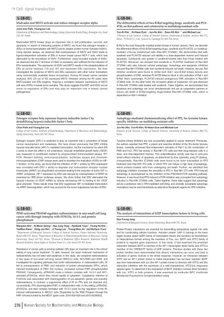
Cell: signal transductionL-18-01hSalvador and MST2 activate and reduce estrogen receptor alphaJoon-woo Park, Young-joo LeeDepartment of Bioscience and Biotechnology, Sejong University Kunja-Dong, Kwangjin-Gu, Seoul143-747, KoreaMammalian MST2 kinase plays an important role in cell proliferation, survival, andapoptosis. In search of interacting proteins of MST2, we found that estrogen receptor α(ERα) co-immunoprecipitates with MST2 and its adaptor protein human Salvador (hSAV).Using reporter assays, we observed that overexpression of MST2 and hSAV leads toligand-independent activation of ERαin human breast cancer MCF-7 cells, which wasattenuated by the knockdown of hSAV. Furthermore, using truncated mutants of hSAV,we observed that the C terminus of hSAV is necessary and sufficient for the induction ofERαtransactivation. The expression of hSAV and MST2 results in the phosphorylation ofERαat serine residues <strong>11</strong>8 and 167 and represses ERαexpression. We theninvestigated the incidence of MST2 and ERαexpression with other tumor biomarkersusing commercially available tissue microarrays. Among 40 breast cancer samplesanalyzed, 60% (24 out of 40) expressed MST2. Nineteen among the 40 cases wereMST2-positive and ERα-negative, implying a correlation between expressions of MST2with loss of ERαin breast tumor samples. This study suggests that MST and hSAV act asnovel co-regulators of ERαand may play an important role in breast cancerpathogenesis.L-18-04The differential effects of two B-Raf targeting drugs, sorafenib and PLX-4720, on Raf pathway and cytotoxicity in multidrug-resistant cellsYun-Ki Kim¹, Ki-Hwan Eum¹, Jun-Ho Ahn¹, Soon Kil Ahn¹ , ²and Michael Lee¹¹Division of Life Sciences, College of Natural Sciences, University of Incheon, Incheon 406-772,Korea, ²YOUAI Co., Ltd., Suwon-Si, Gyeonggi-Do 443-766, KoreaB-Raf is the most frequently mutated protein kinase in human cancers. Here, we describethe differential effects of two B-Raf targeting drugs, sorafenib and PLX4720, on multidrugresistantv-Ha-ras transformed cells (Ras-NIH 3T3/Mdr). We demonstrate that bothsorafenib and PLX4720 reduced cell viability in a dose-dependent manner and inducedapoptosis. Cytotoxicity was greater in sorafenib-treated cells than those treated withPLX4720. Moreover, we showed that sorafenib or PLX4720 treatment of Ras-NIH3T3/Mdr cells also induced cellular autophagy. Autophagy and apoptosis inhibitionrescued Ras-NIH 3T3/Mdr cells from sorafenib-induced cell death. However, the pan Rafinhibitor sorafenib were found to inhibit both Raf-1 and B-Raf kinase activity andphosphorylation of ERK, whereas PLX4720 failed to block in vitro activation of Raf-1 andB-Raf. More surprisingly, PLX4720 induced endogenous ERK activation in Ras-NIH3T3/Mdr cells. On the other hand, the increased uptake of rhodamine <strong>12</strong>3 was observedin Ras-NIH 3T3/Mdr cells treated with sorafenib. Taken together, we demonstrated thatapoptosis and autophagy can occur simultaneously and act as cooperative partners toinduce cell death in B-Raf targeting drugs-treated Ras-NIH 3T3/Mdr cells, which isdependent on Raf-1 inhibition.L-18-02Estrogen receptor beta represses hypoxia inducible factor-1 bydestabilizing hypoxia inducible factor-1 betaChoa Park and YoungJoo LeeCollege of Life Science, Institute of Biotechnology, Department of Bioscience and Biotechnology,Sejong University, Seoul 143-747, KoreaEstrogen receptor (ER) βis predicted to play an important role in prevention of breastcancer development and metastasis. We have shown previously that ERβinhibitshypoxia inducible factor (HIF)-1αmediated transcription, but the mechanism by which ERβworks to exert this effect is not understood. Vascular endothelial growth factor (VEGF)was measured in conditioned medium by enzyme-linked immunosorbent assays. RT-PCR, Western blotting, immunoprecipitation, luciferase assays and chromatinimmunoprecipitation (ChIP) assays were used to ascertain the implication of ERβon HIF-1 function. In this study, we found that the inhibition of HIF-1 activity by ERβexpressionwas correlated with ERβ’s ability to degrade aryl hydrocarbon receptor nucleartranslocator (ARNT) via ubiquitination processes leading to the reduction of active HIF-1α/ARNT complexes. HIF-1 repression by ERβwas rescued by overexpression of ARNT asexamined by HRE-driven luciferase assays. We show further that ERβattenuated thehypoxic induction of VEGF mRNA by directly decreasing HIF-1αbinding to the VEGFgene promoter. These results show that ERβsuppresses HIF-1α-mediated transcriptionvia ARNT downregulation, which may account for the tumor suppressive function of ERβ.L-18-05Autophagy-mediated chemosensitizing effect of PP2, Src tyrosine kinasespecific inhibitor, on multidrug resistant cellsJun-Ho Ahn, Yun-Ki Kim, Ki-Hwan Eum and Michael LeeDivision of Life Sciences, College of Natural Sciences, University of Incheon, Incheon 406-772,KoreaTyrosine kinase inhibitors are now successfully applied to cancer treatment. Previously,the authors reported that PP2, a potent and selective inhibitor of the Src-family tyrosinekinase, markedly enhanced Ras-independent activation of Raf-1 by the combination ofPMA and H2O2. PP2 has activity in Ras-NIH 3T3 cells and their drug-resistant cells in adose- and time-dependent manner. This antiproliferative activity was due to cell-cyclearrest without induction of apoptosis, as determined by flow cytometry using PI staining.Unexpectedly, Ras-NIH 3T3/Mdr cells were found to be more susceptible to PP2treatment than Ras-NIH 3T3 cells, in which PP2 can induce a high level of autophagy,which protects treated cells from undergoing cell death. Further, we identified thatautophagy inhibition increases the sensitivity to PP2 in Ras-NIH 3T3 cells. PP2-inducedautophagy is accompanied by the inhibition of the PI3K/Akt/mTOR signaling pathway.However, it was found that PP2-induced mTOR inhibition was uncoupled from autophagyinduction in Ras-NIH 3T3/Mdr cells. Taken together, these data suggest that autophagyserves a protective role in PP2-mediated cell killing, and clinically acceptable autophagymodulators may be used beneficially as adjunctive therapeutic agents for SFK inhibitors.L-18-03PIM1-activated PRAS40 regulates radioresistance in non-small cell lungcancer cells through interplay with FOXO3a, 14-3-3, and proteinphosphatasesWanyeon Kim¹, Ki Moon Seong³, Hee Jung Yang¹, HyeSook Youn², Young Ha Kim¹,TaeWoo Kwon¹, Dong Jun Kim¹, Ji Young Lee¹, Young-Woo Jin³and BuHyun Youn¹¹Department of Biological Sciences, College of Natural Sciences, Pusan National University,Busan 609-735, Korea, ²Department of Bioscience & Biotechnology/Institute of Bioscience, SejongUniversity, Seoul 143-747, Korea, ³Division of Radiation Effect Research, Radiation HealthResearch Institute, Korea Hydro & Nuclear Power Co., Ltd, Seoul 132-703, KoreaResistance of cancer cells to ionizing radiation (IR) plays an important role in the clinicalsetting of lung cancer treatment. To date, however, the exact molecular mechanism ofradiosensitivity has not been well explained. In this study, we compared radioresistancein two types of non-small cell lung cancer (NSCLC) cells, NCI-H460 and A549, andinvestigated the signaling pathways that confer radioresistance. In radioresistant cells, IRled to overexpression of PIM1 and reduction of protein phosphatases (PPs), whichinduced translocation of PIM1 into nucleus. Increased nuclear PIM1 phosphorylatedPRAS40. Consequently, pPRAS40 made a trimeric complex with 14-3-3 and AKTactivatedpFOXO3a, which then moved rapidly to cytoplasm. Cytoplasmic retention ofFOXO3a was associated with downregulation of pro-apoptotic genes and possiblyradioresistance. In contrast, a suppressive effect of IR on PPs was not detected and,concomitantly, PPs downregulated PIM1 in radiosensitive cells. In this setting, pPRAS40,pFOXO3a, and their complex formation with 14-3-3 could be key regulators of the IRinducedradioresistance in NSCLC cells. [Supported by the R&D Program through theNRF of Korea funded by the MEST (grant code: 20<strong>10</strong>-<strong>00</strong>21920 and 20<strong>10</strong>-<strong>00</strong>29553)]L-18-06The analysis of interactions of bZIP transcription factors in living cellsHye-Young JangDepartment of Biological Sciences, Kosin University, Busan 606-701, KoreaProtein-Protein interactions are essential for transmitting extracellular signals into cellsand for coordinating cellular functions. Activator protein 1(AP-1) belongs to the basicregion leucine zipper (bZIP) family of transcription factors ans functions as homodimersor heterodimers formed among the members of Fos, Jun, BATF and ATF2 family ofproteins to regulate gene expression. In this study, it had examined the predictedinteraction between BATF-a member of the AP-1 transcription factor family and ATF2-amember of the CREB/ATF family of bZIP proteins. Previous studies with these twoprotein families have demonstrated that dimeric interactions can occur and lead toactivation of genes involves in the stress response. However, an interaction betweenATF2 and an AP-1 protein known to inhibit transcription has not been reported. BATFalso form heterodimers with Jun (the AP-1 protein known to dimerize with ATF2) and thisinteraction interferes with the expression of a well-characterized, ATF2/Jun responsivereporter gene. To determine if the mechanism of BATF inhibition involves dimer formationwith Jun, ATF2 or both proteins, it was examined by multicolor BiFC (multicolorBimolecular Fluorescence Complementation) assay.276 Korean Society for Biochemistry and Molecular Biology



![No 기ê´ëª
(êµë¬¸) ëíì ì íë²í¸ ì¹ì£¼ì ì·¨ê¸í목[ì문] ë¶ì¤ë²í¸ 1 ...](https://img.yumpu.com/32795694/1/190x135/no-eeeeu-e-eii-i-iei-ii-1-4-i-ieiecie-eiei-1-.jpg?quality=85)
