11:10-12:00, Rm 103
11:10-12:00, Rm 103
11:10-12:00, Rm 103
You also want an ePaper? Increase the reach of your titles
YUMPU automatically turns print PDFs into web optimized ePapers that Google loves.
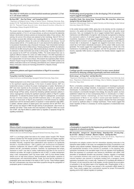
Development and regenerationE-17-01The effect of vitrification on mitochondrial membrane potential (ΔΨm)in 2- cell mouse embryosBo-Hyun KIM¹ , ², Kyu-Tae Chang³and Young-Kug CHOO¹¹Department of Biological Science, College of Natural Sciences, Wonkwang University, Iksan,Jeonbuk 570-749, ²Eulji Medi-Bio Research Institute, Eulji University, Seong Nam, Gyeong Gi-Do461-713, ³National Primate Research Center, Korea Research Institute of Bioscience &Biotechnology, Ochang, Chungbuk 363-883, KoreaThe present study was designed to investigate the effect of vitrification on mitochondrialmembrane potential (ΔΨm) in 2- cell mouse embryos, as well as to document the relationshipbetween mitochondrial membrane potential and developmental ability of those embryos. Afterwarming, the survival of the embryos was assessed by their morphology. Most (91-<strong>10</strong>0 %) ofthe embryos recovered after vitrification were morphologically normal. The rates of blastocystdid not differ significantly between the control and vitrified-warmed 2-cell embryos. Two-cellembryos stained with the ΔΨm-sensitive dye (JC-1) were observed for the distribution of hightolow-polarized mitochondria using a fluorescent microscope. Fresh mitochondrial membranepotential was similar to that of vitrified embryos. Following staining by DCHFDA, the distributionof H2O2 did not differ among the groups. Mitochondrial staining by rhodamin <strong>12</strong>3 showed thatthe distribution of viable mitochondria were similar in all the groups. The DNA fragmentation ofwas assessed by the dUTP nick end-labeling (TUNEL). The results showed there was nosignificant difference between the control and vitrified embryos in the DNA fragmentation ofblastocyst nuclei. In conclusion, vitrification seems to be an effective, easy and rapid method forthe cryopreservation of 2-cell mouse embryos. [This research was supported by Basic ScienceResearch Program through the National Research Foundation of Korea (NRF) funded by theMinistry of Education Science and Technology (20<strong>10</strong>-<strong>00</strong>22316) and a research grant from theMinistry of Education, Science and Technology (KGC540<strong>10</strong><strong>11</strong>), Republic of korea]E-17-02Expression patterns and signal modulations of Rgs19 in secondarypalateYoung Rae Ji and Zae Young RyooSchool of Life Science and Biotechnology, Kyungpook National University, Daegu, KoreaPalatal development is one of the crucial events in craniofacial morphogenesis, accordingto the significant signaling pathway. In the fusion of palatal shelves, EMT is afundamental process to achieve the proper morphogenesis of palate. Mechanisms ofEMT have been reported as the processes of migration, apoptosis or general EMTthrough the modulations through various signalling molecules. Rgs19, known as a RGSfamily through GTPase activity, showed the interesting epithelial expression patterns invarious organogenesis including the significant expression patterns of Rgs19 in palataldevelopment. To evaluate the precise function of Rgs19 in palatogenesis, we employedthe loss of function studies using AS-ODN treatments while in vitro palate organcultivations. Knock-down of Rgs19 using treatments of AS-ODN showed the retardedpalatal fusion with the decreased patterns of apoptosis in mesial epithelium edge (MEE).In addition, alteration patterns of related genes were examined with the qRT-PCR. AndEMT process was delayed in MEE throught staining of pancytokeratin, which known asepithelial cell marker. These results show that expression of Rgs19 in MEE has crucialrole of EMT, also Rgs19 affects to palatal fusion by regulation of apoptosis through thesignalling modulations.E-17-04Proliferating neural progenitors in the developing CNS of zebrafishrequire jagged2 and jagged1bJung-Woo Gwak, Hee Jeong Kong, Young-Ki Bae, Min Jung Kim, Jehee Lee,Jeong-Ho Park and Sang-Yeob YeoDepartment of Biotechnology, Division of Applied Chemistry and Biotechnology, Hanbat NationalUniversity, Daejeon 305-719, KoreaIn the central nervous system (CNS), giving rise to the diversity and the complexity ofneurons is the spatial and temporal differentiation of neural stem cells and/or neuralprecursors. Here, we investigated the role of Jagged mediated Notch signaling in themaintenance and differentiation of progenitor cells during late neurogenesis by analyzingthe expression patterns of zebrafish jagged homologues, and by injecting theirmorpholinos. Expression of both jagged2 and jagged1b mRNA in the CNS suggestedthat they might be involved in control of differentiating neural progenitors in which they areinvolved later in development. In Jagged2 and Jagged1b knock-down embryos, theoverall rate of cell division dramatically decreased, and the ectopic VeMe neurons weregenerated. The results suggest that Jagged-Notch signaling plays a critical role in themaintenance of proliferating neural precursors, and that the generation of late-bornneurons, especially VeMe neurons, is regulated by the interplay between Jagged2 andJagged1b.E-17-05Cartilage specific overexpression of Nkx3.2 in mice causes skeletaldwarfism by delaying cartilage hypertrophy and bone ossificationDa-Un Jeong¹, Je-Yong Choi²and Dae-Won Kim¹¹Department of Biochemistry, College of Life Science and Biotechnology, Yonsei University, Seoul,Korea, ²Department of Biochemistry and Cell Biology, School of Medicine, Kyungpook NationalUniversity, Daegu, KoreaNkx3.2, a homeobox containing protein, is initially expressed in chondrogenic precursorcells, and later during cartilage maturation, its expression is diminished in hypertrophicchondrocytes. To understand an in vivo role of Nkx3.2 during cartilage development, wehave employed a cre-inducible transgenic mice strategy. By using this system, we haveestablished multiple lines of mice overexpressing Nkx3.2 in chondrocytes in a Col2a1-Cre-dependent manner. Interestingly, we have found that cartilage-specificoverexpression of Nkx3.2 in vivo results in severe post-natal dwarfism in mostendochondral elements. In addition, various analyses indicated that these phenotypesare caused by significant delays in cartilage hypertrophy. These results suggest thatproper regulation of Nkx3.2 is necessary for normal progression of endochondral bonedevelopment.E-17-03Effects of Jazf1 overexpression on mouse cardiac functionKi Beom Bae and Zae Young RyooSchool of Life Science and Biotechnology, Kyungpook National University, Daegu, KoreaJAZF1 is a zinc-finger protein that binds to the nuclear orphan receptor TR4. Recentevidence indicates that TR4 receptor functions both as a positive and negative regulatorof transcription, but the role of JAZF1 in transcriptional mechanisms has not beenelucidated. Recently, congenital heart malformations were reported to be significantlyelevated in neurofibromatosis 1 (NF1) patients with chromosomal microdeletionsyndrome. Furthermore, the Joined to JAZF1 is highly expressed in the hearts of adultpatients with NF1 microdeletion syndrome. Therefore, we hypothesized that ectopicexpression of JAZF1 may lead to cardiac malformations that allow for the survival ofnewborns and adults. We sought to elucidate the role of JAZF1 in cardiac developmentusing a Jazf1 overexpression (Jazf1-Tg) mouse model. Jazf1-Tg mice showed cardiacdefects such as high blood pressure, electrocardiogram abnormalities, apoptosis ofcardiomyocytes, and mitochondrial defects. Finally, we found that the expressions ofproapoptotic genes were elevated in the hearts of Jazf1-Tg mice. These findings suggestthat Jazf1 overexpression may induce heart failure symptoms through the upregulation ofproapoptotic genes in cardiomyocytes.E-17-06α-Syntrophin is required for the hepatocyte growth factor-inducedmigration of cultured myoblastsMin-Jeong Kim¹, Jeong-A Lim¹, Sung-Ho Hwang¹, Sang-Hyun Kim¹, Ki-Jung Jang¹,Su-Jin Choi¹, Stanley C. Froehner², Marvin E. Adams²and Hye-Sun Kim¹¹Department of Biological Science, Ajou University, Suwon 443-749 Korea, ²Department ofPhysiology and Biophysics, University of Washington, WA 98195-7290 USASyntrophins are adaptor proteins that link intracellular signaling molecules to thedystrophin based scaffold. In this study, we investigated the function of syntrophins in cellmigration using cultured myoblasts. Hepatocyte growth factor (HGF) stimulates migrationand lamellipodial formation of the cultured C2 myoblasts. In the migrating cells,syntrophins concentrate in the rear-lateral region of the cell, opposite of the lamellipodia.When the expression of α-syntrophin was reduced by transfection with siRNA, HGFstimulation of lamellipodia formation was prevented. Likewise, migration of myoblastsfrom α-syntrophin knockout mice could not be stimulated by HGF. However, HGFinduced migration was restored in myoblasts isolated from a transgenic mouseexpressing α-syntrophin only in muscle cells. Treatment of C2 myoblasts with inhibitors ofPI3-kinase, not only reduced the rate of cell migration, but also impaired the accumulationof syntrophins in the rear-lateral region of the migrating cells. Phosphorylation of Akt, adownstream effecter of PI3-kinase, was reduced in the α-syntrophin siRNA treated C2cells. These results suggest that α-syntrophinis required for proper PI3-kinase/Aktsignaling and for HGF-induced migration of myoblasts.230 Korean Society for Biochemistry and Molecular Biology




![No 기ê´ëª
(êµë¬¸) ëíì ì íë²í¸ ì¹ì£¼ì ì·¨ê¸í목[ì문] ë¶ì¤ë²í¸ 1 ...](https://img.yumpu.com/32795694/1/190x135/no-eeeeu-e-eii-i-iei-ii-1-4-i-ieiecie-eiei-1-.jpg?quality=85)
