11:10-12:00, Rm 103
11:10-12:00, Rm 103
11:10-12:00, Rm 103
You also want an ePaper? Increase the reach of your titles
YUMPU automatically turns print PDFs into web optimized ePapers that Google loves.
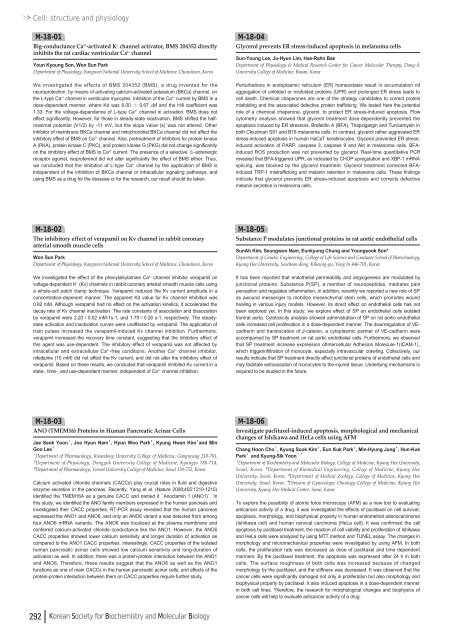
Cell: structure and physiologyM-18-01Big-conductance Ca 2+ -activated K + channel activator, BMS 204352 directlyinhibits the rat cardiac ventricular Ca 2+ channelYoun Kyoung Son, Won Sun ParkDepartment of Physiology, Kangwon National University School of Medicine, Chuncheon, KoreaWe investigated the effects of BMS 204352 (BMS), a drug invented for theneuroprotection, by means of activating calcium-activated potassium (BKCa) channel, onthe L-type Ca 2+ channel in ventricular myocytes. Inhibition of the Ca 2+ current by BMS in adose-dependent manner, where Kd was 6.<strong>00</strong> ± 0.67 μM and the Hill coefficient was1.33. For the voltage-dependence of L-type Ca 2+ channel in activation, BMS does notaffect significantly. However, for those in steady-state inactivation, BMS shifted the halfmaximalpotential (V1/2) by -<strong>11</strong> mV, but the slope value (k) was not altered. Otherinhibitor of membrane BKCa channel and mitochondrial BKCa channel did not affect theinhibitory effect of BMS on Ca 2+ channel. Also, pretreatment of inhibitors for protein kinaseA (PKA), protein kinase C (PKC), and protein kinase G (PKG) did not change significantlyon the inhibitory effect of BMS to Ca 2+ current. The presence of a selective -adrenergicreceptor agonist, isoproterenol did not alter significantly the effect of BMS either. Thus,we concluded that the inhibition of L-type Ca 2+ channel by the application of BMS isindependent of the inhibition of BKCa channel or intracellular signaling pathways, andusing BMS as a drug for the desease or for the research, our result should be taken.M-18-04Glycerol prevents ER stress-induced apoptosis in melanoma cellsSun-Young Lee, Ju-Hyun Lim, Hae-Rahn BaeDepartment of Physiology & Medical Research Center for Cancer Molecular Therapy, Dong-AUniversity College of Medicine, Busan, KoreaPerturbations in endoplasmic reticulum (ER) homeostasis result in accumulation ndaggregation of unfolded or misfolded proteins (UPR) and prolonged ER stress leads tocell death. Chemical chaperones are one of the strategy candidates to correct proteinmisfolding and the associated defective protein trafficking. We tested here the potentialrole of a chemical chaperone, glycerol, to protect ER stress-induced apoptosis. Flowcytometry analysis showed that glycerol treatment dose-dependently prevented theapoptosis induced by ER stressors, Brefeldin A (BFA), Thapsigargin and Tunicamysin inboth Cloudman S91 and B16 melanoma cells. In contrast, glycerol rather aggravated ERstress-induced apoptosis in human HaCaT keratinocytes. Glycerol prevented ER stressinducedactivation of PARP, caspase 3, caspase 9 and Akt in melanoma cells. BFAinducedROS production was not prevented by glycerol. Real-time quantitative PCRrevealed that BFA-triggered UPR, as indicated by CHOP upregulation and XBP-1 mRNAsplicing, was blocked by the glycerol treatment. Glycerol treatment corrected BFAinducedTRP-1 mistrafficking and melanin retention in melanoma cells. These findingsindicate that glycerol prevents ER stress-induced apoptosis and corrects defectivemelanin secretion in melanoma cells.M-18-02The inhibitory effect of verapamil on Kv channel in rabbit coronaryarterial smooth muscle cellsWon Sun ParkDepartment of Physiology, Kangwon National University School of Medicine, Chuncheon, KoreaWe investigated the effect of the phenylalkylamine Ca 2+ channel inhibitor verapamil onvoltage-dependent K + (Kv) channels in rabbit coronary arterial smooth muscle cells usinga whole-cell patch clamp technique. Verapamil reduced the Kv current amplitude in aconcentration-depenent manner. The apparent Kd value for Kv channel inhibition was0.82 mM. Although verapamil had no effect on the activation kinetics, it accelerated thedecay rate of Kv channel inactivation. The rate constants of association and dissociationby verapamil were 2.20±0.02 mM-1s-1, and 1.79±0.26 s-1, respectively. The steadystateactivation and inactivation curves were unaffected by verapamil. The application oftrain pulses increased the verapamil-induced Kv channel inhibition. Furthermore,verapamil increased the recovery time constant, suggesting that the inhibitory effect ofthis agent was use-dependent. The inhibitory effect of verapamil was not affected byintracellular and extracellular Ca 2+ -free conditions. Another Ca 2+ channel inhibitor,nifedipine (<strong>10</strong> mM) did not affect the Kv current, and did not alter the inhibitory effect ofverapamil. Based on these results, we concluded that verapamil inhibited Kv current in astate-, time-, and use-dependent manner, independent of Ca 2+ channel inhibition.M-18-05Substance P modulates junctional proteins in rat aortic endothelial cellsSunAh Kim, Seungwoo Nam, Eunkyung Chung and Youngsook Son*Department of Genetic Engineering, College of Life Science and Graduate School of Biotechnology,Kyung Hee University, Seocheon-dong, Kiheung-gu, Yong In 446-701, KoreaIt has been reported that endothelial permeability and angiogenesis are modulated byjunctional proteins. Substance P(SP), a member of neuropeptides, mediates painperception and regulates inflammation. In addition, recently we reported a new role of SPas awound messenger to mobilize mesenchymal stem cells, which promotes woundhealing in various injury models. However, its direct effect on endothelial cells has notbeen explored yet. In this study, we explore effect of SP on endothelial cells isolatedfromrat aorta. Cytotoxicity analysis showed administration of SP on rat aortic endothelialcells increased cell proliferation in a dose-dependent manner. The downregulation of VEcadherinand translocation of β-catenin, a cytoplasmic partner of VE-cadherin wereaccompanied by SP treatment on rat aortic endothelial cells. Furthermore, we observedthat SP treatment increase expression ofIntercellular Adhesion Molecule-1(ICAM-1),which triggerinfiltration of monocyte, especially intravascular crawling. Collectively, ourresults indicate that SP treatment directly affect junctional proteins of endothelial cells andmay facilitate extravasation of monocytes to the injured tissue. Underlying mechanisms isrequired to be studied in the future.M-18-03ANO (TMEM16) Proteins in Human Pancreatic Acinar CellsJae Seok Yoon¹, Joo Hyun Nam², Hyun Woo Park³, Kyung Hwan Kim³and MinGoo Lee³¹Department of Pharmacology, Kwandong University College of Medicine, Gangneung 2<strong>10</strong>-701,²Department of Physiology, Dongguk University College of Medicine, Kyungju 780-714,³Department of Pharmacology, Yonsei University College of Medicine, Seoul <strong>12</strong>0-752, KoreaCalcium activated chloride channels (CACCs) play crucial roles in fluid and digestiveenzyme secretion in the pancreas, Recently, Yang et al. (Nature 2<strong>00</strong>8;455:<strong>12</strong><strong>10</strong>-<strong>12</strong>15)identified the TMEM16A as a genuine CACC and named it ‘Anoctamin 1 (ANO1)’. Inthis study, we identified the ANO family members expressed in the human pancreas andinvestigated their CACC properties. RT-PCR assay revealed that the human pancreasexpressed the ANO1 and ANO6, and only an ANO6 variant a was detected from amongfour ANO6 mRNA variants. The ANO6 was localized at the plasma membrane andconferred calcium-activated chloride conductance like the ANO1. However, the ANO6CACC properties showed lower calcium sensitivity and longer duration of activation ascompared to the ANO1 CACC properties. Interestingly, CACC properties of the isolatedhuman pancreatic acinar cells showed low calcium sensitivity and long-duration ofactivation as well. In addition, there was a protein-protein interaction between the ANO1and ANO6. Therefore, these results suggest that the ANO6 as well as the ANO1functions as one of main CACCs in the human pancreatic acinar cells, and effects of theprotein-protein interaction between them on CACC properties require further study.M-18-06Investigate paclitaxel-induced apoptosis, morphological and mechanicalchanges of Ishikawa and HeLa cells using AFMChang Hoon Cho¹, Kyung Sook Kim², Eun Kuk Park³, Min-Hyung Jung⁴, Hun-KukPark²and Kyung-Sik Yoon¹ , ¹Department of Biochemistry and Molecular Biology, College of Medicine, Kyung Hee University,Seoul, Korea, ²Department of Biomedical Engineering, College of Medicine, Kyung HeeUniversity, Seoul, Korea, ³Department of Medical Zoology, College of Medicine, Kyung HeeUniversity, Seoul, Korea, ⁴Division of Gynecologic Oncology College of Medicine, Kyung HeeUniversity, Kyung Hee Medical Center, Seoul, KoreaTo explore the possibility of atomic force microscopy (AFM) as a new tool to evaluatinganticancer activity of a drug, it was investigated the effects of paclitaxel on cell survival,apoptosis, morphology, and biophysical property in human endometrial adenocarcinoma(Ishikawa cell) and human cervical carcinoma (HeLa cell). It was confirmed the cellapoptosis by paclitaxel treatment, the reaction of cell viability and proliferation of Ishikawaand HeLa cells were analyzed by using MTT method and TUNEL assay. The changes inmorphology and micromechanical properties were investigated by using AFM. In bothcells, the proliferation rate was decreased as dose of paclitaxel and time dependentmanners. By the paclitaxel treatment, the apoptosis was expressed after 24 h in bothcells. The surface roughness of both cells was increased because of changedmorphology by the paclitaxel, and the stiffness was decreased. It was observed that thecancer cells were significantly damaged not only in proliferation but also morphology andbiophysical property by paclitaxel. It also induced apoptosis in a dose-dependent mannerin both cell lines. Therefore, the research for morphological changes and biophysics ofcancer cells will help to evaluate anticancer activity of a drug.292 Korean Society for Biochemistry and Molecular Biology



![No 기ê´ëª
(êµë¬¸) ëíì ì íë²í¸ ì¹ì£¼ì ì·¨ê¸í목[ì문] ë¶ì¤ë²í¸ 1 ...](https://img.yumpu.com/32795694/1/190x135/no-eeeeu-e-eii-i-iei-ii-1-4-i-ieiecie-eiei-1-.jpg?quality=85)


