11:10-12:00, Rm 103
11:10-12:00, Rm 103
11:10-12:00, Rm 103
You also want an ePaper? Increase the reach of your titles
YUMPU automatically turns print PDFs into web optimized ePapers that Google loves.
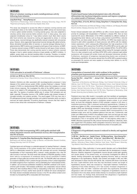
NeuroscienceH-17-61Effect of voluntary running on matrix metalloproteinase activityfollowing brain ischemiaJung-Seok Hong¹ , ², Seong-Ryong Lee¹¹Department of Pharmacology, School of Medicine, Keimyung University, Daegu, 704-701²Department of Emergency, Ulsan University Hospital, Ulsan, KoreaThis study was designed to access the effect of voluntary running exercise andimmobilization stress on the matrix metalloproteinase (MMP) activity and neuronal injuryin focal or global cerebral ischemia. In running exercise group, mice were subjected tovoluntary activity wheel exercise before ischemic insult. In stress group, mice weresubjected immobilization stress for 7 days before ischemic insult. Brain infarct volumeafter focal ischemia was reduced by running and aggravated by stress. In runningexercise group, neuronal damage was significantly reduced compared with non-exercisegroup. However, stress aggravated hippocampal neuronal damage after global ischemia.Stress diminished beneficial effect of exercise on neuronal damage following focal orglobal ischemia. MMP-9 activity was increased by both types of brain ischemia, not MMP-2. Exercise reduced increase of MMP-9 activity induced by both types of brain ischemia.These data demonstrate that running exercise seems to reduce ischemic neuronaldamage in focal or global cerebral ischemia at least partially via MMP-9 inhibition. Inaddition, severe stress can diminish beneficial effect of exercise. Therefore, to maintainthe beneficial effect of exercise, it is important to reduce or avoid stress-inducing events.H-17-64Protein-based human induced pluripotent stem cells efficientlydifferentiate into functional dopamine neurons and improve symptomsof a rodent model of Parkinson’s diseaseYong-Hee Rhee, Ji-Yun Ko, Mi-Yoon Chang, Sang-Hoon Yi, Kwang-Soo Kim, Sang-Hun LeeDepartment of Biochemistry and Molecular Biology, College of Medicine, and Hanyang BiomedicalResearch Institute, Hanyang University, Seoul 133-791, KoreaHuman induced pluripotent stem cells (hiPSCs) can offer a human disease model andpromise for autologus cell transplantation in Parkinson’s disease (PD). Here we haveexamined DA neuron differentiation of the hiPSCs established by different methods ofreprogramming factor delivery such as those using lentiviral (Lenti-hiPSCs), retroviral(Retro-iPSCs), and direct protein delivery (Pro-hiPSCs). All hiPSC lines tested could beinduced to yield uniform populations of neural precursor cells (NPCs) with the midbrainmarker expressions, which subsequently differentiated into high proportions of DAneurons. However, NPCs derived from Pro-hiPSCs (Pro-hiPSC-NPCs) are the safer andstable DA neuronal source over those of viral vector-mediated hiPSCs. Pro-hiPSC-NPCswere highly expandable for at least 8 passages without losing DA neurogenic potential.This is in a clear contrast to Lenti- and Retro-hiPSC NPCs which underwent rapid cellularsenescence with increased p53 protein levels and decreased telomerase activities.Furthermore, expression of exogenous reprogramming factors was continued indifferentiated Lenti-hiPSCs. DA neurons derived from Pro-hiPSC-NPCs exerted functionsas presynaptic DA neurons and were capable of reversing motor deficits in a rat PDmodel upon transplantation.H-17-62TLR3 activation in rat model of Parkinson’s diseaseJin-Suk Lee, Mi-Ra Lee, Won Gil ChoDepartment of Anatomy, Wonju College of Medicine, Yonsei University, Wonju 220-701, KoreaSystemic infections are often associated with neurodegenerative processes in manydiseases of the central nervous system (CNS), including Parkinson’s disease. Toll-likereceptor 3 (TLR3) is a double-stranded RNA (dsRNA) sensor that mediates an anti-viralinnate immune response. We investigated the effect of the dsRNA injection in areasknown to be related to PD pathogenesis on rat including striatum (ST) and substantianigra (SN). The injection of synthetic dsRNA poly IC evokes TLR3 activation and celldeath, especially TH-positive neurons in SN. dsRNA injection also showed increase ofmicroglial activation and cytokine secretion. This result suggest TLR3 may have role inPD pathogenesis through microglial activation and cytokine expression which can lead toneuronal cell death especially TH-positive dopaminergic neurons that their death isknown to have critical role in development of Parkinson’s disease.H-17-63Nurr1 and ca-Raf overexpressing NPCs yield grafts enriched withmature dopamine neurons and functional recovery after transplantationinto PD rat brainsSang-Hoon Yi, Yong-Hee Rhee, Boe-Kyoung Kim, Sang-Hun LeeBiochemistry and Molecular Biology, College of Medicine, and Biomedical Reasearch Insititute,Hanyang University, Seoul 133-791, KoreaCombined gene and cell therapy is a potential approach for treating Parkinson’s disease(PD). Forced expression of the nuclear receptor Nurr1 drives neural precursor cells(NPCs) to differentiate towards DA neurons with promoted cell survival. Intrastriataltransplantation of Nurr1-expressing NPCs, however, mostly generates only a few DAcells survived in host brain. We show herein that activation of Raf-ERK signaling in thesedonor cells by co-expression of constitutive active (ca)-raf contributes to faithfulgeneration of large grafts fully enriched with DA cells. The Raf-ERK effect was achievedby increased availability of Nurr1 proteins through prevention of Nurr1 protein degradationand stable exogenous Nurr1 expression, and by potentiating Nurr1-transcriptional activity.In addition, cell survival and proliferation mediated by Raf-ERK contribute to DA neuronenrichedgraft formation. Raf-mediated proliferation was substantially reduced withoutaltering the other beneficial effects of Raf by manipulating ca-Raf dose, cell density andby co-expression of Foxa2, another factor specific for midbrain DA neuron development.We finally showed remarkable behavioral recovery in the PD rats without evident tumorformation by transplanting NPCs manipulated with those factors.H-17-65Upregulation of neuronal nitric oxide synthase in the peripherypromotes pain hypersensitivity after peripheral nerve injuryKyung Hwa Kim¹, Jong-Il Kim¹ , ², Jeong A Han³, Myoung-Ae Choe⁴ , *, and Jung-Hyuck Ahn 5, *¹Department of Biochemistry and Molecular Biology, Seoul National University College ofMedicine, Seoul <strong>11</strong>0-799, Korea, ²Department of Biomedical Sciences, Seoul National UniversityGraduate School, Seoul <strong>11</strong>0-799, Korea, ³Department of Biochemistry and Molecular Biology,Kangwon National University College of Medicine, Chuncheon 2<strong>00</strong>-701, Korea, ⁴College ofNursing, Seoul National University, Seoul <strong>11</strong>0-799, Korea, 5 Department of Biochemistry, EwhaWomans University School of Medicine, Seoul 158-7<strong>10</strong>, KoreaPeripheral nerve injury often results in neuropathic pain that manifests as hyperalgesia,and allodynia. Several studies suggest a functional role for neuronal nitric oxide synthase(nNOS) on neuropathic pain, but such a contribution remains unclear. In our currentstudy, we found that intraplantar injection of NOS substrate L-arginine or NO donor 3-morpholino-synonimine (SIN-1) produced mechanical hypersensitivity. Following L5spinal nerve ligation (L5 SNL), immunoreactivity for nNOS in the ipsilateral L5 dorsal rootganglion (DRG) was dramatically increased in mainly small- and medium-sized neuronsand non-neuronal cells. L5 SNL caused increased nNOS immunoreactivity in theipsilateral sciatic nerve and the ipsilateral glabrous hind paw skin. Furthermore, totalprotein and mRNA levels of nNOS in the ipsilateral sciatic nerve and hind paw skin weremarkedly upregulated following nerve injury. Intraplantar injection of NOS inhibitor 7-nitroindazole (7-NI) or non-specific NOS inhibitor L-N G -nitro-arginine methyl ester (L-NAME) effectively suppressed the SNL-induced mechanical allodynia. Collectively, thesedata suggest that in the periphery nNOS upregulation induced by nerve injury contributesto mechanical hypersensitivity during the maintenance phase of neuropathic pain.H-17-66Ciliogenesis of hypothalamic neuron is reduced in obesity and regulatedby leptinYu Mi Han¹, Sae Bom Lee¹, Hyukki Chang³, Gil Myoung Kang¹, Hyun-Kyong Kim¹,So Young Gil¹, Joong Yeol Park², Ki Up Lee², Hyuk Whan Koh⁴, Min-Seon Kim²¹Appetite Regulation Laboratory, Asan Institute for Life Science, ²Department of InternalMedicine, University of Ulsan College of Medicine, Seoul, Korea, ³Seoul Women’s University,⁴Age-Related & Brain Diseases Research Center Kyung Hee University, KoreaBackground/Aim: Ciliary dysfunction in human genetic disorders such as Bardet-BiedelSyndrome (BBS) and Alström syndrome is associated with the development of obesity.Moreover, defective cilogenesis in hypothalamic neurons disrupts central leptin signalingand causes obesity. In the present study, we conversely investigated the effects ofobesity, feeding manipulation and appetite regulating hormone, leptin on neuronalciliogenesis in the hypothalamus. Results: Cilia numbers and lengths in ARC and VMHwere decreased in obese mice fed with high-fat- diet, compared with those in low-fat-dietfed mice. Leptin-deficient ob/ob mice also had decreased number and length ofhypothalamic neurons, which was reversed by 7 days-leptin treatment. Furthermore,regaining of food intake following a 24 h fast acutely increased ciliogenesis inhypothalamic ARC and VMH. Treatment with leptin also significantly stimulatedciliogenesis in both cell models. Conclusions: Ciliogenesis of hypothalamic neurons isdecreased in the in two obese animal models whereas it is increased by leptin treatmentand food intake. Our findings firstly demonstrate that ciliogenesis in hypothalamicneurons is dynamically regulated by metabolic conditions.254 Korean Society for Biochemistry and Molecular Biology


![No 기ê´ëª
(êµë¬¸) ëíì ì íë²í¸ ì¹ì£¼ì ì·¨ê¸í목[ì문] ë¶ì¤ë²í¸ 1 ...](https://img.yumpu.com/32795694/1/190x135/no-eeeeu-e-eii-i-iei-ii-1-4-i-ieiecie-eiei-1-.jpg?quality=85)
