11:10-12:00, Rm 103
11:10-12:00, Rm 103
11:10-12:00, Rm 103
You also want an ePaper? Increase the reach of your titles
YUMPU automatically turns print PDFs into web optimized ePapers that Google loves.
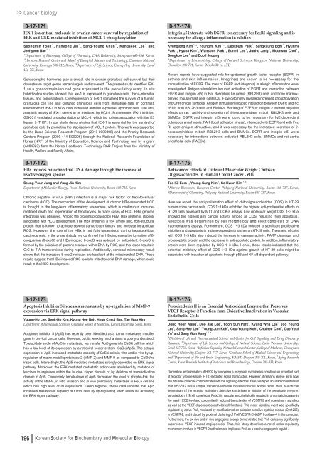
Cancer biologyB-17-171IEX-1 is a critical molecule in ovarian cancer survival by regulation ofERK and GSK-mediated inhibition of MCL-1 phosphorylationSeongmin Yoon¹, Hanyong Jin¹, Sang-Young Chun², Kangseok Lee³andJeehyeon Bae¹ , *¹Department of Pharmacy, College of Pharmacy, CHA University, Seongnam 463-836, Korea,²Hormone Research Center and School of Biological Sciences and Technology, Chonnam NationalUniversity, Kwangju 5<strong>00</strong>-7<strong>12</strong>, Korea, ³Department of Life Science, Chung-Ang University, Seoul156-756, KoreaGonadotrophic hormones play a crucial role in ovarian granulosa cell survival but theirdownstream target genes remain largely undiscovered. The present study identified IEX-1 as a gonadotropin-induced gene expressed in the preovulatory ovary. In situhybridization studies showed that Iex-1 is expressed in granulosa cells, theca-interstitialtissues, and corpus luteum. Overexpression of IEX-1 stimulated the survival of a humangranulosa cell line and cultured granulosa cells from immature rats. In contrast,knockdown of IEX-1 in KGN cells increased annexin V-positive, apoptotic cells. The antiapoptoticactivity of IEX-1 seems to be mediated by MCL-1. Furthermore, IEX-1 inhibitedGSK-3-mediated phosphorylation of MCL-1, which led to less association with the E3ligase -TrCP. In our study demonstrates that IEX-1 is essential for the survival ofgranulosa cells by promoting the stabilization of MCL-1 protein. This work was supportedby the Basic Science Research Program (20<strong>10</strong>-<strong>00</strong>09498) and the Priority ResearchCenters Program (2<strong>00</strong>9-414-E<strong>00</strong><strong>00</strong>6) through the National Research Foundation ofKorea (NRF) of the Ministry of Education, Science and Technology and by a grant(A084923) from the Korea Healthcare Technology R&D Project from the Ministry ofHealth, Welfare and Family Affairs.B-17-172HBx induces mitochondrial DNA damage through the increase ofreactive oxygen speciesSeung-Youn Jung and Yung-Jin KimDepartment of Molecular Biology, Pusan National University, Busan 609-735, KoreaChronic hepatitis B virus (HBV) infection is a major risk factor for hepatocellularcarcinoma (HCC). The mechanism of the development of chronic HBV-associated HCCis thought to the long-term inflammatory responses, which is continuous immunemediateddeath and regeneration of hepatocytes. In many cases of HCC, HBV genomeintegration was observed. Among the proteins produced by HBV, HBx protein is stronglyassociated with HCC development. The HBx protein is a 154 amino acid, non-structuralprotein that is known to activate several transcription factors and increase intracellularROS. However, the role of the HBx is not fully understood during hepatocellularcarcinogenesis. In the present study, we confirmed that HBx increases the formation of 8-oxoguanine (8-oxoG) and HBx-induced 8-oxoG was reduced by antioxidant. 8-oxoG isformed by the oxidation of guanine residues within DNA by ROS, and this lesion results inG:C to T:A transversions during replication. Additionally, confocal microscopy resultshows that the increased 8-oxoG residues are localized at the mitochondrial DNA. Theseresults suggest that HBx-induced ROS leads to mitochondrial DNA damage, which couldresult in the HCC development.B-17-174Integrin α5 interacts with EGFR, is necessary for FcεRI signaling and isnecessary for allergic inflammation in relationKyungjong Kim¹ , *, Youngmi Kim¹ , *, Deokbum Park¹, Sangkyung Eom¹, HyunmiPark¹, Hyuna Kim¹, Wansoon Park¹, Eunmi Lee¹, Junho Jang¹, Moonsun Choi¹,Songkoo Lee²and Dooil Jeoung¹¹Department of Biochemistry, College of Natural Sciences, Kangwon National University,Chunchon 2<strong>00</strong>-701, Korea, ²Mushville co. LTDRecent reports have suggested role for epidermal growth factor receptor (EGFR) inasthma and skin inflammation. Integrin(s) are known to be necessary for thetransactivation of EGFR. The roles of EGFR and integrin(s) in allergic inflammation wereinvestigated. Antigen stimulation induced activation of EGFR and interaction betweenEGFR and integrin α(5) in Rat Basophilic Leukemia (RBL2H3) cells and bone marrowderivedmouse mast cells (BMMCs). Flow cytometry revealed increased phosphorylationof EGFR on cell surfaces. Antigen stimulation induced interaction between EGFR and FcεRI in both RBL2H3 cells and BMMCs. Blocking of EGFR or integrin αexerted negativeeffects on rac1 activity and secretion of β-hexosaminidase in both RBL2H3 cells andBMMCs. EGFR and integrin α(5) were found to be necessary for IgE-dependentcutaneous anaphylaxis. FAK (focal adhesion kinase), interacted with EGFR and with FcεRI upon antigen stimulation, and it was necessary for the increased secretion of β-hexosaminidase in both RBL2H3 cells and BMMCs. EGFR and integrin α(5) werenecessary for interactions between activated RBL2H3 cells, BMMCs and rat aorticendothelial cells (RAECs).B-17-175Anti-cancer Effects of Different Molecular Weight ChitosanOligosaccharides in Human Colon Cancer CellsTae-Kil Eom¹, Young-Sang Kim², Se-Kwon Kim¹ , ²¹Marine Bioprocess Research Center, Pukyong National University, Busan 608-737, Korea,²Department of Chemistry, Pukyong National University, Busan 608-737, KoreaHere we report the anti-proliferation effect of chitooligosaccharides (COS) in HT-29human colon cancer cells. COS 1~3 kDa exhibited the highest anti-proliferative effects inHT-29 cells assessed by MTT and CCK-8 assays. Low molecular weight COS 1~3 kDashowed the highest anti cancer activity among all COS, resulting from apoptosis.Apoptosis was determined by cell morphology and electrophoresis of DNAfragmentations assays. Furthermore, COS 1~3 kDa induced a significant proliferativeinhibition and apoptosis in a dose-dependent manner on HT-29 cells. Treatment of cellswith COS 1~3 kDa also induced the increase in caspase activity, PARP cleavage, andpro-apoptotic protein and the decrease in anti-apoptotic protein. In addition, inflammatoryprotein were down-regulated by COS 1~3 kDa. Hence, these results indicated that thepotential inhibitory effect of COS 1~3 kDa against growth of HT-29 cells might beassociated with induction of apoptosis through p53 and NF-κB dependent pathway.B-17-173Apoptosis Inhibitor 5 increases metastasis by up-regulation of MMP-9expression via ERK signal pathwayYoung-Ho Lee, Seok-Ho Kim, Kyung Hee Noh, Hyun Cheol Bae, Tae Woo KimDepartment of Biomedical Sciences, Graduate School of Medicine, Korea University, Seoul, KoreaApoptosis inhibitor 5 (Api5) has recently been identified as a tumor metastasis modifiergene in cervical cancer cells. However, but its working mechanisms is poorly understood.To elucidate a role of Api5 in metastasis, we transfer Api5 gene into CaSki cell line whichhas a low level of its expression by a retroviral vector system (CaSki/Api5). The ectopicexpression of Api5 increased metastatic capacity of CaSki cells in vitro and in vivo by upregulationof matrix metalloproteinase-2 (MMP-2) and MMP-9 as compared to CaSki/noinsert cells. Interestingly, the Api5-mediated metastasis strongly depended on ERK signalpathway. Moreover, the ERK-mediated metastatic action was abolished by mutation ofleucines to arginines within the leucine zipper domain or by deletion of transactivationdomain in Api5. Conversely, knock-down of Api5 decreased the level of phopho-Erk, theactivity of the MMPs, in vitro invasion and in vivo pulmonary metastasis in HeLa cell linewhich has high level of its expression. Taken together, these data indicate that Api5increases metastastic capacity of tumor cells by up-regulating MMP levels via activatingthe ERK signal pathway.B-17-176Peroxiredoxin II is an Essential Antioxidant Enzyme that PreservesVEGF Receptor-2 Function from Oxidative Inactivation in VascularEndothelial CellsDong Hoon Kang 1 , Doo Jae Lee 1 , Yoon Sun Park 1 , Kyung Wha Lee 1 , Joo YoungLee 1 , Sang-Hee Lee 3 , Young Jun Koh 4 , Gou-Young Koh 4 , Chulhee Choi 5 , Dae-YeulYu 6 and Sang Won Kang 1, 2, *¹Division of Life and Pharmaceutical Science and Center for Cell Signaling and Drug DiscoveryResearch, ²Department of Life Science and College of Natural Science, Ewha Womans University,Seoul <strong>12</strong>7-750, Korea, ³Infection Signaling Network Research Center, College of Medicine, ChungnamNational University, Daejeon 301-747, Korea, ⁴Graduate School of Medical Science and Engineeringand 5 Department of Bio and Brain Engineering, KAIST, DaeJeon 305-701, Korea, 6 Aging ResearchCenter, Korea Research Institute of BioScience and Biotechnology, Daejeon 305-333, KoreaGeneration and elimination of H2O2 by endogenous enzymatic machineries constitute an important partof receptor tyrosine kinase (RTK)-mediated signal transduction. However, it remains elusive as to howthis diffusible molecule communicates with the signaling effectors. Here, we report an unanticipated resultthat VEGFR2 has a unique oxidation-sensitive cysteine residue whose redox state is a crucialdeterminant of the receptor activation. Selective knockdown or ablation of the peroxidase enzyme,peroxiredoxin II (PrxII, gene locus Prdx2) in vascular endothelial cells resulted in a dramatic increase inthe basal H2O2 level and concomitantly reduced the activation of VEGFR-2 and downstream signalingas well as the VEGF-dependent endothelial cell functions. This redox signaling event was specificallyregulated by active PrxII, mediated by modification of an oxidation-sensitive cysteine residue (Cys<strong>12</strong>06)in VEGFR-2, and induced by proximal clustering of PrxII/VEGFR-2/NADPH oxidase-4 in the caveolae.Furthermore, the ex vivo and in vivo angiogenic assays demonstrated that PrxII deficiency significantlysuppressed VEGF-induced angiogenesis. Thus, this study describes a novel redox regulatorymechanism involved in VEGFR-2 activation and implicates PrxII as a positive angiogenic regulat .196 Korean Society for Biochemistry and Molecular Biology




![No 기ê´ëª
(êµë¬¸) ëíì ì íë²í¸ ì¹ì£¼ì ì·¨ê¸í목[ì문] ë¶ì¤ë²í¸ 1 ...](https://img.yumpu.com/32795694/1/190x135/no-eeeeu-e-eii-i-iei-ii-1-4-i-ieiecie-eiei-1-.jpg?quality=85)
