11:10-12:00, Rm 103
11:10-12:00, Rm 103
11:10-12:00, Rm 103
Create successful ePaper yourself
Turn your PDF publications into a flip-book with our unique Google optimized e-Paper software.
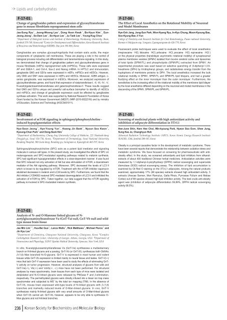
Lipids and carbohydratesF-17-01Change of gangliosides pattern and expression of glycosyltransferasegene in mouse fibroblasts reprogrammed stem cellsJae-Sung Ryu¹, Jeong-Woong Lee², Dong Hoon Kwak¹, Bo-Hyun Kim¹, Eun-Jeong Jeong², So-Dam Lee¹, So-Hyun Lee¹, Ju-Taek Lee¹, Young-Kug Choo¹¹Department of Biological Science and Institute of Biotechnology Wonkwang University, Iksan,Jeonbuk 570-749, Korea and ²Center for Development and Differentiation, Korea Research Instituteof Bioscience and Biotechnology (KRIBB), Dae-jeon 305-806, KoreaGangliosides are complex glycosphingolipids that contain sialic acids, the majorcomponents of cytoplasmic cell membranes. These are playing a role in the control ofbiological process including cell differentiation and tansmembrane signaling. In this study,we demonstrated that change of gangliosides pattern and glycosyltransferase gene inmouse fibroblasts (mEFs) reprogrammed cells (miPSCs). These cells were expressedAP and SSEA-1, and Oct-4, Sox-2 and Nanog mRNA. In HPTLC and immunostain,ganglioside GM3 and GD1a were expressed in mEFs, miPSCs and mESCs. However,only GM2 and GM1 were expressed in mEFs and mESCs. Moreover, A2B5 antigen, c-series ganglioside, was expressed in mESCs. Moreover, we analyzed expression ofglycosyltransferase genes, and found that expression of sialytransferase-I, -II, -III, -IV, -V,N-acetrylgalactosaminetransferase and galactosyltransferas-II. These results suggestthat GM3 and GD1a unique and powerful cell-surface biomarker to identify of mESCsand miPSCs, and change of ganglioside expression could be affected by gangliosdessynthase activation. This work was supported by National Research Foundation of KoreaGrant funded by the Korean Government (MEST) (NRF-20<strong>10</strong>-<strong>00</strong>22316) and by ministryof Education, Science and Technology (KGC540<strong>10</strong><strong>11</strong>).F-17-04The Effect of Local Anesthetics on the Rotational Mobility of Neuronaland Model MembranesHye-Ock Jang, Jong-Sun Park, Won-Hyang Ryu, In-Kyo Chung, Moon-Kyoung Bae,Soo-Kyung Bae, Il YunCollege of Dentistry and Research Institute for Oral Biotechnology, Pusan national University,Beomeo-ri Mulgeum-eup, Yangsan, Gyeongsangnam-do 626-770, KoreaFluorescent probe techniques were used to evaluate the effect of local anesthetics(mepivacaine∙HCl, lidocaine∙HCl, prilocaine∙HCl, procaine∙HCl, ropivacaine∙HCl )on the physical properties (transbilayer asymmetric rotational mobility) of synaptosomalplasma membrane vesicles (SPMV) isolated from bovine cerebral cortex and liposomesof total lipids (SPMVTL) and phospholipids (SPMVPL) extracted from SPMV. Anexperimental procedure was used based on selective quenching of ,6-diphenyl-1,3,5-hexatriene (DPH) by trinitrophenyl groups, and radiationless energy transfer from thetryptophans of membrane proteins to DPH. The local anesthetics increased the bulkrotational mobility in SPMV, SPMVTL and SPMVPL lipid bilayers, and had a greaterfluidizing effect on the inner monolayer than the outer monolayer. Furthermore, thesensitivities to the increasing effect of the rotational mobility of the membrane lipid bilayerby the local anesthetics differed depending on the neuronal and model membranes in thedescending of the SPMV, SPMVPL and SPMVPL.F-17-02Involvement of mTOR signaling in sphingosylphosphorylcholine -induced hypopigmentation effectsHyo-Soon Jeong¹, Hye-Young Yun¹, Kwang Jin Baek¹, Nyoun Soo Kwon¹,Kyoung-Chan Park²and Dong-Seok Kim¹¹Department of Biochemistry, Chung-Ang University College of Medicine, 221 Heukseok-dongDongjak-gu, Seoul 156-756, Korea, ²Department of Dermatology, Seoul National UniversityBundang Hospital, 3<strong>00</strong> Gumi-dong, Bundang-gu, Seongnam-si, Kyoungki-do 463-707, KoreaSphingosylphosphorylcholine (SPC) acts as a potent lipid mediator and signalingmolecule in various cell types. In the present study, we investigated the effects of SPC onmelanogenesis and SPC-modulated signaling pathways related to melanin synthesis.SPC had significant hypopigmentation effects in a dose-dependent manner. It was foundthat SPC induced not only activation of Akt but also stimulation of mTOR, a downstreammediator of the Akt signaling pathway. Moreover, SPC decreased the levels of LC3 IIwhich is known to be regulated by mTOR. Treatment with the mTOR inhibitor rapamycinabolished decreases in melanin and LC3II levels by SPC. Furthermore, we found that theAkt inhibitor LY294<strong>00</strong>2 restored SPC-mediated downregulation of LC3 II and inhibited theactivation of mTOR by SPC. Taken together, our data suggest that the mTOR signalingpathway is involved in SPC-modulated melanin synthesis.F-17-05Screening of medicinal plants with high antioxidant activity andinhibition of adipocyte differentiation in 3T3-L1Hee-June Shin, Nam Hee Choi, Min-kyoung Park, Hyeon Soo Eom, Uhee Jung,Sung Kee Jo, Changhyun RohAdvanced Radiation Technology Institute (ARTI), Korea Atomic Energy Research Institute(KAERI), <strong>12</strong>66, Jeonbuk 580-185, KoreaObesity is a principal causative factor in the development of metabolic syndrome. Therehave been several reports that demonstrate the relationship between oxidative stress andmetabolic syndrome. We have focused on screening for pharmaceuticals with antiobesityeffect. In this study, we screened antioxidants and lipid inhibitors from ethanolextractsof about 4<strong>00</strong> traditional Chinese herbal medicines. Antioxidative activities weremeasured by 1,1-diphenyl-2-picrylhydrazyl (DPPH) radical scavenging and superoxidedismutase (SOD) radical scavenging assay. The inhibition of lipid accumulation isexamined by Oil Red O staining on the 3T3-L1 adipocytes. Among the natural productsexamined, approximately 17% (68 species) extracts showed high antioxidant activity. 5extracts (Arecae Semen, Mori Ramulus, Galla Rhois, Pyrrosiae Folium and MeliaeCortex) out of 68 species showed high lipid inhibition activity. The best crude anti-obesityagent was (inhibition of adipocyte differentiation 59.89%, DPPH radical scavengingactivity 90.5%).F-17-03Analysis of N- and O-Mannose linked glycans of N-acetylglucosaminyltransferase Va (GnT-Va) null, GnT-Vb null and wildtype mouse brain tissueJae Min Lim¹ , ², Hua-Bei Guo², Lance Wells², Rick Matthews³, Michael Pierce²andJin Kyu Lee²¹Department of Chemistry, Changwon National University, Changwon, Korea ²ComplexCarbohydrate Research Center, University of Georgia, Athens, Georgia, USA ³Department ofNeuroscience and Physiology, SUNY Upstate Medical University, Syracuse, New York, USAIn vitro, N-acetylglucosaminyltransferase Va (GnT-Va) synthesizes a multiantennarybranch on N-linked glycans and a paralog, GnT-Vb (or GnT-IX), synthesizes both GlcNAcβ-(1,6) Man branched N-/O-glycans. GnT-V is expressed in most human and rodenttissues while GnT-Vb expression is limited mainly to neural tissue and testes. GnT-V(-/-)mice that lack GnT-V expression have been used to study the effects of eliminating GnT-V activity on tumor progression. However, structural analyses of glycans from wild andGnT-Va(-/-), Vb(-/-) and VaVb(-/-, -/-) mice have not been performed. For structuralanalyses by mass spectrometry, brain tissues from each type of mice were isolated anddelipidated and N-/O-linked glycans were released by PNGase F and β-elimination,respectively. The permethylated glycans were directly infused into a linear ion trap massspectrometer and subjected to MS nby the total ion mapping (TIM). In the absence ofGnT-Vb, mouse brain expressed wild-type levels of N-linked glycans with β-(1,6)branches and markedly reduced levels of O-Man-linked glycans. In vivo, GnT-Vsynthesizes mainly N-linked glycans with very small amounts of O-Man-linked glycanswhen GnT-Vb cannot act. GnT-Vb, however, appears to be only able to synthesize O-Man glycans and not N-linked branches.236 Korean Society for Biochemistry and Molecular Biology



![No 기ê´ëª
(êµë¬¸) ëíì ì íë²í¸ ì¹ì£¼ì ì·¨ê¸í목[ì문] ë¶ì¤ë²í¸ 1 ...](https://img.yumpu.com/32795694/1/190x135/no-eeeeu-e-eii-i-iei-ii-1-4-i-ieiecie-eiei-1-.jpg?quality=85)
