11:10-12:00, Rm 103
11:10-12:00, Rm 103
11:10-12:00, Rm 103
Create successful ePaper yourself
Turn your PDF publications into a flip-book with our unique Google optimized e-Paper software.
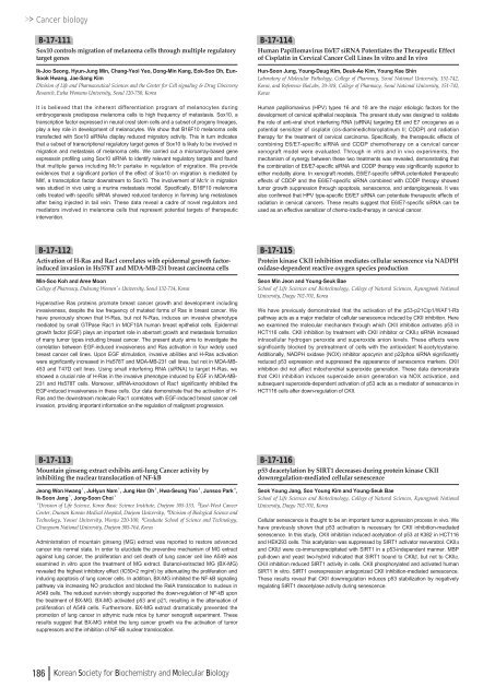
Cancer biologyB-17-<strong>11</strong>1Sox<strong>10</strong> controls migration of melanoma cells through multiple regulatorytarget genesIk-Joo Seong, Hyun-Jung Min, Chang-Yeol Yeo, Dong-Min Kang, Eok-Soo Oh, Eun-Sook Hwang, Jae-Sang KimDivision of Life and Pharmaceutical Sciences and the Center for Cell signaling & Drug DiscoveryResearch, Ewha Womans University, Seoul <strong>12</strong>0-750, KoreaIt is believed that the inherent differentiation program of melanocytes duringembryogenesis predispose melanoma cells to high frequency of metastasis. Sox<strong>10</strong>, atranscription factor expressed in neural crest stem cells and a subset of progeny lineages,play a key role in development of melanocytes. We show that B16F<strong>10</strong> melanoma cellstransfected with Sox<strong>10</strong> siRNAs display reduced migratory activity. This in turn indicatesthat a subset of transcriptional regulatory target genes of Sox<strong>10</strong> is likely to be involved inmigration and metastasis of melanoma cells. We carried out a microarray-based geneexpresssin profiling using Sox<strong>10</strong> siRNA to identify relevant regulatory targets and foundthat multiple genes including Mc1r partake in regulation of migration. We provideevidences that a significant portion of the effect of Sox<strong>10</strong> on migration is mediated byMitf, a transcription factor downstream to Sox<strong>10</strong>. The involvement of Mc1r in migrationwas studied in vivo using a murine metastasis model. Specifically, B16F<strong>10</strong> melanomacells treated with specific siRNA showed reduced tendency in forming lung metastasesafter being injected in tail vein. These data reveal a cadre of novel regulators andmediators involved in melanoma cells that represent potential targets of therapeuticintervention.B-17-<strong>11</strong>4Human Papillomavirus E6/E7 siRNA Potentiates the Therapeutic Effectof Cisplatin in Cervical Cancer Cell Lines In vitro and In vivoHun-Soon Jung, Young-Deug Kim, Deuk-Ae Kim, Young Kee ShinLaboratory of Molecular Pathology, College of Pharmacy, Seoul National University, 151-742,Korea, and Reference BioLabs, 20-318, College of Pharmacy, Seoul National University, 151-742,KoreaHuman papillomavirus (HPV) types 16 and 18 are the major etiologic factors for thedevelopment of cervical epithelial neoplasia. The present study was designed to validatethe role of anti-viral short interfering RNA (siRNA) targeting E6 and E7 oncogenes as apotential sensitizer of cisplatin (cis-diaminedichloroplatinum II; CDDP) and radiationtherapy for the treatment of cervical carcinoma. Specifically, the therapeutic effects ofcombining E6/E7-specific siRNA and CDDP chemotherapy on a cervical cancerxenograft model were evaluated. Through in vitro and in vivo experiments, themechanism of synergy between these two treatments was revealed, demonstrating thatthe combination of E6/E7-specific siRNA and CDDP therapy was significantly superior toeither modality alone. In xenograft models, E6/E7-specific siRNA potentiated therapeuticeffects of CDDP and the E6/E7-specific siRNA combined with CDDP therapy showedtumor growth suppression through apoptosis, senescence, and antiangiogenesis. It wasalso confirmed that HPV type-specific E6/E7 siRNA can potentiate therapeutic effects ofradiation in cervical cancers. These results suggest that E6/E7-specific siRNA can beused as an effective sensitizer of chemo-/radio-therapy in cervical cancer.B-17-<strong>11</strong>2Activation of H-Ras and Rac1 correlates with epidermal growth factorinducedinvasion in Hs578T and MDA-MB-231 breast carcinoma cellsMin-Soo Koh and Aree MoonCollege of Pharmacy, Duksung Women’s University, Seoul 132-714, KoreaHyperactive Ras proteins promote breast cancer growth and development includinginvasiveness, despite the low frequency of mutated forms of Ras in breast cancer. Wehave previously shown that H-Ras, but not N-Ras, induces an invasive phenotypemediated by small GTPase Rac1 in MCF<strong>10</strong>A human breast epithelial cells. Epidermalgrowth factor (EGF) plays an important role in aberrant growth and metastasis formationof many tumor types including breast cancer. The present study aims to investigate thecorrelation between EGF-induced invasiveness and Ras activation in four widely usedbreast cancer cell lines. Upon EGF stimulation, invasive abilities and H-Ras activationwere significantly increased in Hs578T and MDA-MB-231 cell lines, but not in MDA-MB-453 and T47D cell lines. Using small interfering RNA (siRNA) to target H-Ras, weshowed a crucial role of H-Ras in the invasive phenotype induced by EGF in MDA-MB-231 and Hs578T cells. Moreover, siRNA-knockdown of Rac1 significantly inhibited theEGF-induced invasiveness in these cells. Our data demonstrate that the activation of H-Ras and the downstream molecule Rac1 correlates with EGF-induced breast cancer cellinvasion, providing important information on the regulation of malignant progression.B-17-<strong>11</strong>5Protein kinase CKII inhibition mediates cellular senescence via NADPHoxidase-dependent reactive oxygen species productionSeon Min Jeon and Young-Seuk BaeSchool of Life Sciences and Biotechnology, College of Natural Sciences, Kyungpook NationalUniversity, Daegu 702-701, KoreaWe have previously demonstrated that the activation of the p53-p21Cip1/WAF1-Rbpathway acts as a major mediator of cellular senescence induced by CKII inhibition. Herewe examined the molecular mechanism through which CKII inhibition activates p53 inHCT<strong>11</strong>6 cells. CKII inhibition by treatment with CKII inhibitor or CKIIαsiRNA increasedintracellular hydrogen peroxide and superoxide anion levels. These effects weresignificantly blocked by pretreatment of cells with the antioxidant N-acetylcysteine.Additionally, NADPH oxidase (NOX) inhibitor apocynin and p22phox siRNA significantlyreduced p53 expression and suppressed the appearance of senescence markers. CKIIinhibition did not affect mitochondrial superoxide generation. These data demonstratethat CKII inhibition induces superoxide anion generation via NOX activation, andsubsequent superoxide-dependent activation of p53 acts as a mediator of senescence inHCT<strong>11</strong>6 cells after down-regulation of CKII.B-17-<strong>11</strong>3Mountain ginseng extract exhibits anti-lung Cancer activity byinhibiting the nuclear translocation of NF-kBJeong Won Hwang¹, JuHyun Nam¹, Jung Han Oh², Hwa-Seung Yoo³, Junsoo Park⁴,Ik-Soon Jang¹, Jong-Soon Choi¹¹Division of Life Science, Korea Basic Science Institute, Daejeon 305-333, ²East-West CancerCenter, Dunsan Korean Medical Hospital, Daejeon University, ³Division of Biological Science andTechnology, Yonsei University, Wonju 220-<strong>10</strong>0, ⁴Graduate School of Science and Technology,Chungnam National University, Daejeon 305-764, KoreaAdministration of mountain ginseng (MG) extract was reported to restore advancedcancer into normal state. In order to elucidate the preventive mechanism of MG extractagainst lung cancer, the proliferation and cell death of lung cancer cell line A549 wasexamined in vitro upon the treatment of MG extract. Butanol-extracted MG (BX-MG)revealed the highest inhibitory effect (IC50=2 mg/ml) by attenuating the proliferation andinducing apoptosis of lung cancer cells. In addition, BX-MG inhibited the NF-kB signalingpathway via increasing NO production and blocked the RelA translocation to nucleus inA549 cells. The reduced survivin strongly supported the down-regulation of NF-kB uponthe treatment of BX-MG. BX-MG activated p53 and p21, resulting in the attenuation ofproliferation of A549 cells. Furthermore, BX-MG extract dramatically prevented thepromotion of lung cancer in athymic nude mice by tumor xenograft experiment. Theseresults suggest that BX-MG inhibit the lung cancer growth via the activation of tumorsuppressors and the inhibition of NF-kB nuclear translocation.B-17-<strong>11</strong>6p53 deacetylation by SIRT1 decreases during protein kinase CKIIdownregulation-mediated cellular senescenceSeok Young Jang, Soo Young Kim and Young-Seuk BaeSchool of Life Sciences and Biotechnology, College of Natural Sciences, Kyungpook NationalUniversity, Daegu 702-701, KoreaCellular senescence is thought to be an important tumor suppression process in vivo. Wehave previously shown that p53 activation is necessary for CKII inhibition-mediatedsenescence. In this study, CKII inhibition induced acetylation of p53 at K382 in HCT<strong>11</strong>6and HEK293 cells. This acetylation was suppressed by SIRT1 activator resveratrol. CKIIαand CKIIβwere co-immunoprecipitated with SIRT1 in a p53-independent manner. MBPpull-down and yeast two-hybrid indicated that SIRT1 bound to CKIIβ, but not to CKIIα.CKII inhibition reduced SIRT1 activity in cells. CKII phosphorylated and activated humanSIRT1 in vitro. SIRT1 overexpression antagonized CKII inhibition-mediated senescence.These results reveal that CKII downregulation induces p53 stabilization by negativelyregulating SIRT1 deacetylase activity during senescence.186 Korean Society for Biochemistry and Molecular Biology



![No 기ê´ëª
(êµë¬¸) ëíì ì íë²í¸ ì¹ì£¼ì ì·¨ê¸í목[ì문] ë¶ì¤ë²í¸ 1 ...](https://img.yumpu.com/32795694/1/190x135/no-eeeeu-e-eii-i-iei-ii-1-4-i-ieiecie-eiei-1-.jpg?quality=85)
