11:10-12:00, Rm 103
11:10-12:00, Rm 103
11:10-12:00, Rm 103
Create successful ePaper yourself
Turn your PDF publications into a flip-book with our unique Google optimized e-Paper software.
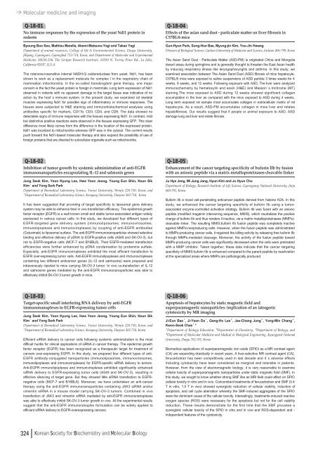
Molecular medicine and imagingQ-18-01No immune responses by the expression of the yeast Ndi1 protein inrodentsByoung Boo Seo, Mathieu Marella, Akemi Matsuno-Yagi and Takao YagiDepartment of animal resources, College of life & Environmental Science, Daegu University,Jillyang, Gyeongsan, Gyeongbuk 7<strong>12</strong>-714, Korea, and Department of Molecular and ExperimentalMedicine, MEM-256, The Scripps Research Institute, <strong>10</strong>550 N. Torrey Pines Rd., La Jolla,California 92037, U.S.A.The rotenone-insensitive internal NADH-Q oxidoreductase from yeast, Ndi1, has beenshown to work as a replacement molecule for complex I in the respiratory chain ofmammalian mitochondria. In the so-called transkingdom gene therapy, one majorconcern is the fact the yeast protein is foreign in mammals. Long term expression of Ndi1observed in rodents with no apparent damage to the target tissue was indicative of noaction by the host’s immune system. In the present study, we examined rat skeletalmuscles expressing Ndi1 for possible sign of inflammatory or immune responses. Thetissues were subjected to H&E staining and immunohistochemical analyses usingantibodies specific for markers, CD<strong>11</strong>b, CD3, CD4, and CD8. The data showed nodetectable signs of immune responses with the tissues expressing Ndi1. In contrast, mildbut distinctive positive reactions were observed in the tissues expressing GFP. This cleardifference most likely comes from the difference in the location of the expressed protein.Ndi1 was localized to mitochondria whereas GFP was in the cytosol. The current resultspush forward the Ndi1-based molecular therapy and also expand the possibility of use offoreign proteins that are directed to subcellular organelle such as mitochondria.Q-18-04Effects of the asian sand dust - particulate matter on liver fibrosis inC57BL/6 miceGun-Hyun Park, Sung-Hun Bae, Myung-jin Kim, You-Jin HwangDivision of Biological Science, Gachon University of Medicine and Science, Incheon 406-799, KoreaThe Asian Sand Dust - Particulate Matter (ASD-PM) is originated China and Mongoliadesert areas during springtime and is generally thought to threaten the East Asian healthby inducing respiratory illness like laryngopharyngitis and asthma. In this study, weexamined association between The Asian Sand Dust (ASD) fibrosis of mice hepatocyte.C57BL/6 mice were exposed to saline suspensions of ASD particle 3 times weeks for 4weeks, 8 weeks, and <strong>12</strong> weeks. Following exposure with ASD, The liver were analyzedimmunochemistry by hematoxylin and eosin (H&E) and Masson’s trichrome (MT)staining.The mice exposed to ASD during <strong>12</strong> weeks showed significant collagenaccumulation in the liver as compared with the mice exposed to ASD during 4 weeks.Long term exposed rat sample most accumulate collagen in extracellular matrix of rathepatocyte. As a result, ASD-PM accumulates collagen in mice liver and relateshepatofibrosis. Our results suggest that if people or animal exposure to ASD, ASDdamage lung and liver and relate fibrosis.Q-18-02Inhibition of tumor growth by systemic administration of anti-EGFRimmunonanoparticles encapsulating IL-<strong>12</strong> and salmosin genesJung Seok Kim, Yeon Kyung Lee, Hwa Yeon Jeong, Young Eun Shin, Keun SikKim¹and Yong Serk ParkDepartment of Biomedical Laboratory Science, Yonsei University, Wonju 220-7<strong>10</strong>, Korea, and¹Department of Biomedical Laboratory Science, Konyang University, Daejeon 302-718, KoreaIt has been suggested that providing of target specificity to liposomal gene deliverysystem may be able to enhance their in vivo transfection efficiency. The epidermal growthfactor receptor (EGFR) is a well known small and stable tumor-associated antigen widelyexpressed in various cancer cells. In this study, we developed four different types ofEGFR-targeted gene delivery system (immunoliposomes, immunovirosomes,immunolipoplexes and immunoviroplexes) by coupling of anti-EGFR antibodies(Cetuximab) to liposomal surface. The anti-EGFR immunonanoparticles showed selectivebinding and effective delivery of pDNA to EGFR-positive cells (A549 and SK-OV-3), butnot to EGFR-negative cells (MCF-7 and B16BL6). Their EGFR-mediated transfectionefficiencies were further enhanced by pDNA condensation by protamine sulfate.Especially, anti-EGFR immunoviroplexes exhibited the most efficient transfection toEGFR over-expressing tumor cells. Anti-EGFR immunolipoplexes and immunoviroplexescontaining two different anticancer genes (IL-<strong>12</strong> and salmosine) were prepared andintravenously injected to mice carrying SK-OV-3 tumor. In vivo co-transfection of IL-<strong>12</strong>and salmosine genes mediated by the anti-EGFR immunonanoparticles was able toeffectively inhibit SK-OV-3 tumor growth in mice.Q-18-05Enhancement of the cancer targeting specificity of buforin IIb by fusionwith an anionic peptide via a matrix metalloproteinases-cleavable linkerJu Hye Jang, Mi Jung Jang, Hyun Kim and Ju Hyun ChoDepartment of Biology, Research Institute of Life Science, Gyeongsang National University, Jinju660-701, KoreaBuforin IIb a novel cell-penetrating anticancer peptide derived from histone H2A. In thisstudy, we enhanced the cancer targeting specificity of buforin IIb using a tumorassociatedenzyme-controlled activation strategy. Buforin IIb was fused with an anionicpeptide (modified magainin intervening sequence, MMIS), which neutralizes the positivecharge of buforin IIb and thus renders it inactive, via a matrix metalloproteinases (MMPs)-cleavable linker. The resulting MMIS:buforin IIb fusion peptide was completely inactiveagainst MMPs-nonproducing cells. However, when the fusion peptide was administratedto MMPs-producing cancer cells, it regained the killing activity by releasing free buforin IIbthrough MMPs-mediated cleavage. Moreover, the activity of the fusion peptide towardMMPs-producing cancer cells was significantly decreased when the cells were pretreatedwith a MMP inhibitor. Taken together, these data indicate that the cancer targetingspecificity of MMIS:buforin IIb is enhanced compared to the parent peptide by reactivationat the specialized areas where MMPs are pathologically produced.Q-18-03Target-specific small interfering RNA delivery by anti-EGFRimmunonanoplexes to EGFR-expressing tumor cellsJung Seok Kim, Yeon Kyung Lee, Hwa Yeon Jeong, Young Eun Shin, Keun SikKim¹and Yong Serk ParkDepartment of Biomedical Laboratory Science, Yonsei University, Wonju 220-7<strong>10</strong>, Korea, and¹Department of Biomedical Laboratory Science, Konyang University, Daejeon 302-718, KoreaEfficient siRNA delivery to cancer cells following systemic administration is the mostdifficult hurdle for clinical applications of siRNA in cancer therapy. The epidermal growthfactor receptor (EGFR) has been recognized as a therapeutic target for treatment ofcancers over-expressing EGFR. In this study, we prepared four different types of anti-EGFR antibody-conjugated nanoparticles (immunoliposomes, immunovirosomes,immunolipoplexes and immunoviroplexes) for EGFR-specific siRNA delivery to tumors.Anti-EGFR immunolipoplexes and immuniviroplexes exhibited significantly enhancedsiRNA delivery to EGFR-expressing tumor cells (A549 and SK-OV-3), resulting ineffective silencing of target gene. But they showed little siRNA transfection to EGFRnegativecells (MCF-7 and B16BL6). Moreover, we have undertaken an anti-cancertherapy using the anti-EGFR immunonanoparticles containing JAK3 siRNA and/orvimentin siRNA in a mouse model carrying SK-OV-3 tumors. Combined in vivotransfection of JAK3 and vimentin siRNA mediated by anti-EGFR immunonanoplexeswas able to effectively inhibit SK-OV-3 tumor growth in vivo. All the experimental resultssuggest that the anti-EGFR immunonanoplex formulation can be widely applied toefficient siRNA delivery to EGFR-overexpressing cancers.Q-18-06Apoptosis of hepatocytes by static magnetic field andsuperparamagnetic nanoparticles: implication of an iatrogeniccytotoxicity by MR imagingJi-Eun Bae¹, Ji-Yeon Do¹, Gang-Ho Lee², Jae-Chang Jung³, Yong-Min Chang⁴,Kwon-Seok Chae¹ , *¹Department of Biology Education, ²Department of Chemistry, ³Department of Biology, and⁴Department of Molecular Medicine and Medical & Biological Engineering, Kyungpook NationalUniversity, Daegu 702-701, KoreaBiomedical applications of superparamagnetic iron oxide (SPIO) as a MR contrast agent(CA) are expanding drastically in recent years. A liver-selective MR contrast agent (CA),ferucarbotran has been competitively used in last decade and it’s adverse effectsincluding cytotoxicity have been considered as marginal and tolerable in patients.However, from the view of electromagnetic biology, it is very reasonable to examinecellular toxicity of superparamagnetic nanoparticles under static magnetic field (SMF). Inthis study, we sought to know whether strong SMF like as MRI field could affect on SPIOcellular toxicity in vitro and in vivo. Concomitant treatments of ferucarbotran and SMF (0.4T in vitro, 1.5 T in vivo) showed synergistic reduction of cellular viability, induction ofapoptosis, and cell cycle aberration whereby the SMF-induced aggregates of the SPIOwere the dominant cause of the cellular toxicity. Interestingly, treatments-induced reactiveoxygen species (ROS) were necessary for the apoptosis but not for the cell viabilityreduction. These results demonstrate for the first time that the SMF provokes asynergistic cellular toxicity of the SPIO in vitro and in vivo and ROS-dependent and -independent features of the cytotoxicity.324 Korean Society for Biochemistry and Molecular Biology



![No 기ê´ëª
(êµë¬¸) ëíì ì íë²í¸ ì¹ì£¼ì ì·¨ê¸í목[ì문] ë¶ì¤ë²í¸ 1 ...](https://img.yumpu.com/32795694/1/190x135/no-eeeeu-e-eii-i-iei-ii-1-4-i-ieiecie-eiei-1-.jpg?quality=85)
