11:10-12:00, Rm 103
11:10-12:00, Rm 103
11:10-12:00, Rm 103
Create successful ePaper yourself
Turn your PDF publications into a flip-book with our unique Google optimized e-Paper software.
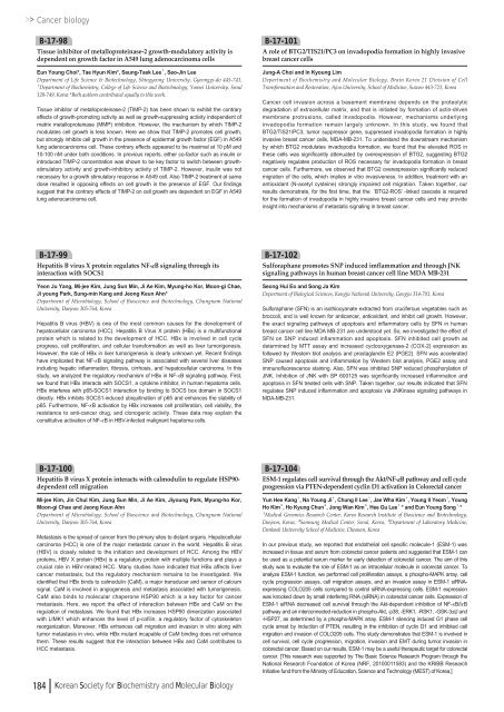
Cancer biologyB-17-98Tissue inhibitor of metalloproteinase-2 growth-modulatory activity isdependent on growth factor in A549 lung adenocarcinoma cellsEun Young Choi*, Tae Hyun Kim*, Seung-Taek Lee¹, Seo-Jin LeeDepartment of Life Science & Biotechnology, Shingyeong University, Gyeonggi-do 445-741,¹Department of Biochemistry, College of Life Science and Biotechnology, Yonsei University, Seoul<strong>12</strong>0-749, Korea *Both authors contributed equally to this work.Tissue inhibitor of metalloproteinase-2 (TIMP-2) has been shown to exhibit the contraryeffects of growth-promoting activity as well as growth-suppressing activity independent ofmatrix metalloproteinase (MMP) inhibition. However, the mechanism by which TIMP-2modulates cell growth is less known. Here we show that TIMP-2 promotes cell growth,but strongly inhibits cell growth in the presence of epidermal growth factor (EGF) in A549lung adenocarcinoma cell. These contrary effects appeared to be maximal at <strong>10</strong> pM and<strong>10</strong>-<strong>10</strong>0 nM under both conditions. In previous reports, either co-factor such as insulin orintroduced TIMP-2 concentration was shown to be key factor to switch between growthstimulatoryactivity and growth-inhibitory activity of TIMP-2. However, insulin was notnecessary for a growth stimulatory response in A549 cell. Also TIMP-2 treatment at samedose resulted in opposing effects on cell growth in the presence of EGF. Our findingssuggest that the contrary effects of TIMP-2 on cell growth are dependent on EGF in A549lung adenocarcinoma cell.B-17-<strong>10</strong>1A role of BTG2/TIS21/PC3 on invadopodia formation in highly invasivebreast cancer cellsJung-A Choi and In Kyoung LimDepartment of Biochemistry and Molecular Biology, Brain Korea 21 Division of CellTransformation and Restoration, Ajou University, School of Medicine, Suwon 443-721, KoreaCancer cell invasion across a basement membrane depends on the proteolyticdegradation of extracellular matrix, and that is initiated by formation of actin-drivenmembrane protrusions, called invadopodia. However, mechanisms underlyinginvadopodia formation remain largely unknown. In this study, we found thatBTG2/TIS21/PC3, tumor suppressor gene, suppressed invadopodia formation in highlyinvasive breast cancer cells, MDA-MB-231. To understand the downstream mechanismby which BTG2 modulates invadopodia formation, we found that the elevated ROS inthese cells was significantly attenuated by overexpression of BTG2, suggesting BTG2negatively regulates production of ROS necessary for invadopodia formation in breastcancer cells. Furthermore, we observed that BTG2 overexpression significantly reducedmigration of the cells, which implies in vitro invasiveness. In addition, treatment with anantioxidant (N-acetyl cysteine) strongly impaired cell migration. Taken together, ourresults demonstrate, for the first time, that the ‘BTG2-ROS’-linked cascade is requiredfor the formation of invadopodia in highly invasive breast cancer cells and may provideinsight into mechanisms of metastatic signaling in breast cancer.B-17-99Hepatitis B virus X protein regulates NF-κB signaling through itsinteraction with SOCS1Yeon Ju Yang, Mi-jee Kim, Jung Sun Min, Ji Ae Kim, Myung-ho Kor, Moon-gi Chae,Ji young Park, Sung-min Kang and Jeong Keun Ahn*Department of Microbiology, School of Bioscience and Biotechnology, Chungnam NationalUniversity, Daejeon 305-764, KoreaHepatitis B virus (HBV) is one of the most common causes for the development ofhepatocellular carcinoma (HCC). Hepatitis B Virus X protein (HBx) is a multifunctionalprotein which is related to the development of HCC. HBx is involved in cell cycleprogress, cell proliferation, and cellular transformation as well as liver tumorigenesis.However, the role of HBx in liver tumorigenesis is clearly unknown yet. Recent findingshave implicated that NF-κB signaling pathway is associated with several liver diseasesincluding hepatic inflammation, fibrosis, cirrhosis, and hepatocellular carcinoma. In thisstudy, we analyzed the regulatory mechanism of HBx in NF-κB signaling pathway. First,we found that HBx interacts with SOCS1, a cytokine inhibitor, in human hepatoma cells.HBx interferes with p65-SOCS1 interaction by binding to SOCS box domain in SOCS1directly. HBx inhibits SOCS1-induced ubiquitination of p65 and enhances the stability ofp65. Furthermore, NF-κB activation by HBx increases cell proliferation, cell viability, theresistance to anti-cancer drug, and clonogenic activity. These data may explain theconstitutive activation of NF-κB in HBV-infected malignant hepatoma cells.B-17-<strong>10</strong>2Sulforaphane promotes SNP induced imflammation and through JNKsignaling pathways in human breast cancer cell line MDA MB-231Seong Hui Eo and Song Ja KimDepartment of Biological Sciences, Kongju National University, Gongju 314-701, KoreaSulforaphane (SFN) is an isothiocyanate extracted from cruciferous vegetables such asbroccoli, and is well known for anticancer, antioxidant, and inhibit cell growth. However,the exact signaling pathways of apoptosis and inflammatory cells by SFN in humanbreast cancer cell line MDA MB-231 are understood yet. So, we investigated the effect ofSFN on SNP induced inflammation and apoptosis. SFN inhibited cell growth asdetermined by MTT assay and increased cyclooxygenase-2 (COX-2) expression asfollowed by Western blot analysis and prostaglandin E2 (PGE2). SFN was acceleratedSNP caused apoptosis and inflammation by Western blot analysis, PGE2 assay andimmunofluorescence staining. Also, SFN was inhibited SNP reduced phosphorylation ofJNK. Inhibition of JNK with SP 6<strong>00</strong><strong>12</strong>5 was significantly increased inflammation andapoptosis in SFN treated cells with SNP. Taken together, our results indicated that SFNregulates SNP induced inflammation and apoptosis via JNKinase signaling pathways inMDA-MB-231.B-17-<strong>10</strong>0Hepatitis B virus X protein interacts with calmodulin to regulate HSP90-dependent cell migrationMi-jee Kim, Jin Chul Kim, Jung Sun Min, Ji Ae Kim, Jiyoung Park, Myung-ho Kor,Moon-gi Chae and Jeong Keun AhnDepartment of Microbiology, School of Bioscience and Biotechnology, Chungnam NationalUniversity, Daejeon 305-764, KoreaMetastasis is the spread of cancer from the primary sites to distant organs. Hepatocellularcarcinoma (HCC) is one of the major metastatic cancer in the world. Hepatitis B virus(HBV) is closely related to the initiation and development of HCC. Among the HBVproteins, HBV X protein (HBx) is a regulatory protein with multiple functions and plays acrucial role in HBV-related HCC. Many studies have indicated that HBx affects livercancer metastasis, but the regulatory mechanism remains to be investigated. Weidentified that HBx binds to calmodulin (CaM), a major transducer and sensor of calciumsignal. CaM is involved in angiogenesis and metastasis associated with tumorigenesis.CaM also binds to molecular chaperone HSP90 which is a key factor for cancermetastasis. Here, we report the effect of interaction between HBx and CaM on theregulation of metastasis. We found that HBx increases HSP90 dimerization associatedwith LIMK1 which enhances the level of p-cofilin, a regulatory factor of cytoskeletonreorganization. Moreover, HBx enhances cell migration and invasion in vitro along withtumor metastasis in vivo, while HBx mutant incapable of CaM binding does not enhancethem. These results suggest that the interaction between HBx and CaM contributes toHCC metastasis.184 Korean Society for Biochemistry and Molecular BiologyB-17-<strong>10</strong>4ESM-1 regulates cell survival through the Akt/NF-κB pathway and cell cycleprogression via PTEN-dependent cyclin D1 activation in Colorectal cancerYun Hee Kang¹, Na Young Ji¹, Chung Il Lee¹, Jae Wha Kim¹, Young Il Yeom¹, YoungHo Kim², Ho Kyung Chun², Jong Wan Kim³, Hee Gu Lee¹ , * and Eun Young Song¹ , *¹Medical Genomics Research Center, Korea Research Institute of Bioscience and Biotechnology,Daejeon, Korea, ²Samsung Medical Center, Seoul, Korea, ³Department of Laboratory Medicine,Dankook University School of Medicine, Cheonan, KoreaIn our previous study, we reported that endothelial cell specific molecule-1 (ESM-1) wasincreased in tissue and serum from colorectal cancer patients and suggested that ESM-1 canbe used as a potential serum marker for early detection of colorectal cancer. The aim of thisstudy was to evaluate the role of ESM-1 as an intracellular molecule in colorectal cancer. Toanalyze ESM-1 function, we performed cell proliferation assays, a phospho-MAPK array, cellcycle progression assays, cell migration assays, and an invasion assay in ESM-1 siRNAexpressingCOLO205 cells compared to control siRNA-expressing cells. ESM-1 expressionwas knocked down by small interfering RNA (siRNA) in colorectal cancer cells. Expression ofESM-1 siRNA decreased cell survival through the Akt-dependent inhibition of NF-κB/IκBpathway and an interconnected reduction in phospho-Akt, -p38, -ERK1, -RSK1, -GSK-3α/βand-HSP27, as determined by a phospho-MAPK array. ESM-1 silencing induced G1 phase cellcycle arrest by induction of PTEN, resulting in the inhibition of cyclin D1 and inhibited cellmigration and invasion of COLO205 cells. This study demonstrates that ESM-1 is involved incell survival, cell cycle progression, migration, invasion and EMT during tumor invasion incolorectal cancer. Based on our results, ESM-1 may be a useful therapeutic target for colorectalcancer. [This research was supported by The Basic Science Research Program through theNational Research Foundation of Korea (NRF, 20<strong>10</strong><strong>00</strong><strong>11</strong>583) and the KRIBB ResearchInitiative fund from the Ministry of Education, Science and Technology (MEST) of Korea.]



![No 기ê´ëª
(êµë¬¸) ëíì ì íë²í¸ ì¹ì£¼ì ì·¨ê¸í목[ì문] ë¶ì¤ë²í¸ 1 ...](https://img.yumpu.com/32795694/1/190x135/no-eeeeu-e-eii-i-iei-ii-1-4-i-ieiecie-eiei-1-.jpg?quality=85)
