11:10-12:00, Rm 103
11:10-12:00, Rm 103
11:10-12:00, Rm 103
Create successful ePaper yourself
Turn your PDF publications into a flip-book with our unique Google optimized e-Paper software.
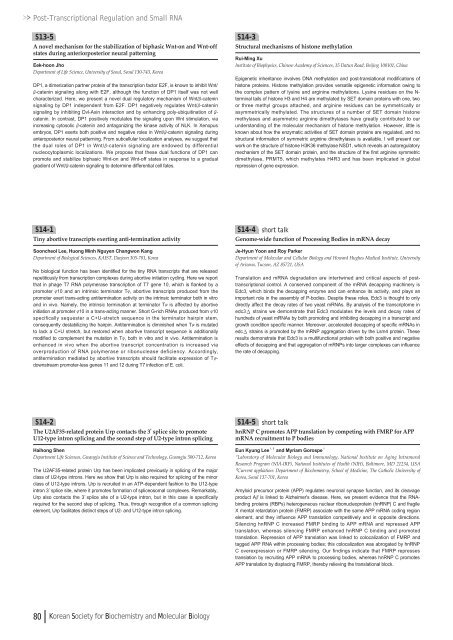
Post-Transcriptional Regulation and Small RNAS13-5A novel mechanism for the stabilization of biphasic Wnt-on and Wnt-offstates during anteriorposterior neural patterningEek-hoon JhoDepartment of Life Science, University of Seoul, Seoul 130-743, KoreaDP1, a dimerization partner protein of the transcription factor E2F, is known to inhibit Wnt/β-catenin signaling along with E2F, although the function of DP1 itself was not wellcharacterized. Here, we present a novel dual regulatory mechanism of Wnt/β-cateninsignaling by DP1 independent from E2F. DP1 negatively regulates Wnt/β-cateninsignaling by inhibiting Dvl-Axin interaction and by enhancing poly-ubiquitination of β-catenin. In contrast, DP1 positively modulates the signaling upon Wnt stimulation, viaincreasing cytosolic β-catenin and antagonizing the kinase activity of NLK. In Xenopusembryos, DP1 exerts both positive and negative roles in Wnt/β-catenin signaling duringanteroposterior neural patterning. From subcellular localization analyses, we suggest thatthe dual roles of DP1 in Wnt/β-catenin signaling are endowed by differentialnucleocytoplasmic localizations. We propose that these dual functions of DP1 canpromote and stabilize biphasic Wnt-on and Wnt-off states in response to a gradualgradient of Wnt/β-catenin signaling to determine differential cell fates.S14-3Structural mechanisms of histone methylationRui-Ming XuInstitute of Biophysics, Chinese Academy of Sciences, 15 Datun Road, Beijing <strong>10</strong>0<strong>10</strong>1, ChinaEpigenetic inheritance involves DNA methylation and post-translational modifications ofhistone proteins. Histone methylation provides versatile epigenetic information owing tothe complex pattern of lysine and arginine methylations. Lysine residues on the N-terminal tails of histone H3 and H4 are methylated by SET domain proteins with one, twoor three methyl groups attached, and arginine residues can be symmetrically orasymmetrically methylated. The structures of a number of SET domain histonemethylases and asymmetric arginine dimethylases have greatly contributed to ourunderstanding of the molecular mechanism of histone methylation. However, little isknown about how the enzymatic activities of SET domain proteins are regulated, and nostructural information of symmetric arginine dimethylases is available. I will present ourwork on the structure of histone H3K36 methylase NSD1, which reveals an autoregulatorymechanism of the SET domain protein, and the structure of the first arginine symmetricdimethylase, PRMT5, which methylates H4R3 and has been implicated in globalrepression of gene expression.S14-1Tiny abortive transcripts exerting anti-termination activitySooncheol Lee, Huong Minh Nguyen Changwon KangDepartment of Biological Sciences, KAIST, Daejeon 305-701, KoreaNo biological function has been identified for the tiny RNA transcripts that are releasedrepetitiously from transcription complexes during abortive initiation cycling. Here we reportthat in phage T7 RNA polymerase transcription of T7 gene <strong>10</strong>, which is flanked by apromoter φ<strong>10</strong> and an intrinsic terminator Tφ, abortive transcripts produced from thepromoter exert trans-acting antitermination activity on the intrinsic terminator both in vitroand in vivo. Namely, the intrinsic termination at terminator Tφis affected by abortiveinitiation at promoter φ<strong>10</strong> in a trans-acting manner. Short G-rich RNAs produced from φ<strong>10</strong>specifically sequester a C+U-stretch sequence in the terminator hairpin stem,consequently destabilizing the hairpin. Antitermination is diminished when Tφis mutatedto lack a C+U stretch, but restored when abortive transcript sequence is additionallymodified to complement the mutation in Tφ, both in vitro and in vivo. Antitermination isenhanced in vivo when the abortive transcript concentration is increased viaoverproduction of RNA polymerase or ribonuclease deficiency. Accordingly,antitermination mediated by abortive transcripts should facilitate expression of Tφdownstreampromoter-less genes <strong>11</strong> and <strong>12</strong> during T7 infection of E. coli.S14-4 short talkGenome-wide function of Processing Bodies in mRNA decayJe-Hyun Yoon and Roy ParkerDepartment of Molecular and Cellular Biology and Howard Hughes Medical Institute, Universityof Arizona, Tucson, AZ 85721, USATranslation and mRNA degradation are intertwined and critical aspects of posttranscriptionalcontrol. A conserved component of the mRNA decapping machinery isEdc3, which binds the decapping enzyme and can enhance its activity, and plays animportant role in the assembly of P-bodies. Despite these roles, Edc3 is thought to onlydirectly affect the decay rates of two yeast mRNAs. By analysis of the transcriptome inedc3 strains we demonstrate that Edc3 modulates the levels and decay rates ofhundreds of yeast mRNAs by both promoting and inhibiting decapping in a transcript andgrowth condition specific manner. Moreover, accelerated decapping of specific mRNAs inedc strains is promoted by the mRNP aggregation driven by the Lsm4 protein. Theseresults demonstrate that Edc3 is a multifunctional protein with both positive and negativeeffects of decapping and that aggregation of mRNPs into larger complexes can influencethe rate of decapping.S14-2The U2AF35-related protein Urp contacts the 3’splice site to promoteU<strong>12</strong>-type intron splicing and the second step of U2-type intron splicingHaihong ShenDepartment Life Sciences, Gwangju Institute of Science and Technology, Gwangju 5<strong>00</strong>-7<strong>12</strong>, KoreaThe U2AF35-related protein Urp has been implicated previously in splicing of the majorclass of U2-type introns. Here we show that Urp is also required for splicing of the minorclass of U<strong>12</strong>-type introns. Urp is recruited in an ATP-dependent fashion to the U<strong>12</strong>-typeintron 3’splice site, where it promotes formation of spliceosomal complexes. Remarkably,Urp also contacts the 3’splice site of a U2-type intron, but in this case is specificallyrequired for the second step of splicing. Thus, through recognition of a common splicingelement, Urp facilitates distinct steps of U2- and U<strong>12</strong>-type intron splicing.S14-5 short talkhnRNP C promotes APP translation by competing with FMRP for APPmRNA recruitment to P bodiesEun Kyung Lee¹ , ²and Myriam Gorospe¹¹Laboratory of Molecular Biology and Immunology, National Institute on Aging IntramuralResearch Program (NIA-IRP), National Institutes of Health (NIH), Baltimore, MD 2<strong>12</strong>34, USA²Current appliation: Department of Biochemistry, School of Medicine, The Catholic University ofKorea, Seoul 137-701, KoreaAmyloid precursor protein (APP) regulates neuronal synapse function, and its cleavageproduct Aβis linked to Alzheimer's disease. Here, we present evidence that the RNAbindingproteins (RBPs) heterogeneous nuclear ribonucleoprotein (hnRNP) C and fragileX mental retardation protein (FMRP) associate with the same APP mRNA coding regionelement, and they influence APP translation competitively and in opposite directions.Silencing hnRNP C increased FMRP binding to APP mRNA and repressed APPtranslation, whereas silencing FMRP enhanced hnRNP C binding and promotedtranslation. Repression of APP translation was linked to colocalization of FMRP andtagged APP RNA within processing bodies; this colocalization was abrogated by hnRNPC overexpression or FMRP silencing. Our findings indicate that FMRP repressestranslation by recruiting APP mRNA to processing bodies, whereas hnRNP C promotesAPP translation by displacing FMRP, thereby relieving the translational block.80 Korean Society for Biochemistry and Molecular Biology



![No 기ê´ëª
(êµë¬¸) ëíì ì íë²í¸ ì¹ì£¼ì ì·¨ê¸í목[ì문] ë¶ì¤ë²í¸ 1 ...](https://img.yumpu.com/32795694/1/190x135/no-eeeeu-e-eii-i-iei-ii-1-4-i-ieiecie-eiei-1-.jpg?quality=85)
