11:10-12:00, Rm 103
11:10-12:00, Rm 103
11:10-12:00, Rm 103
You also want an ePaper? Increase the reach of your titles
YUMPU automatically turns print PDFs into web optimized ePapers that Google loves.
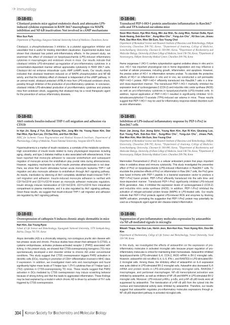
ImmunologyO-18-01Cilostazol protects mice against endotoxin shock and attenuates LPSinducedcytokine expression in RAW 264.7 macrophages via MAPKinhibition and NF-kB inactivation: Not involved in cAMP mechanismsWon Sun ParkDepartment of Physiology, Kangwon National University School of Medicine, Chuncheon, KoreaCilostazol, a phosphodiesterase 3 inhibitor, is a platelet aggregation inhibitor andvasodilator that is useful for treating intermittent claudication. Experimental studies haveshown that cilostazol has potent anti-inflammatory effects. In the present study, weexamined the effect of cilostazol on lipopolysaccharide (LPS)-induced inflammatorycytokines in macrophages and endotoxin shock in mice. Our results indicate thatcilostazol inhibits LPS-stimulated up-regulation of pro-inflammatory cytokines in aconcentration-dependent manner without appreciable cytotoxicity in RAW 264.7 cells.Cilostazol did not enhance intracellular cyclic AMP (cAMP) levels. Our results clearlyindicated that cilostazol treatment reduced on of MAPK phosphorylation and NF-kBacivity, and that the inhibitory effect of cilostazol is independent of the cAMP pathway. Inan animal model, cilostazol protected c57BL/6 mice from LPS-induced endotoxin shock,possibly through inhibition of the production of pro-inflammatory cytokines. In conclusion,cilostazol inhibits LPS-stimulated production of pro-inflammatory cytokines and protectsmice from endotoxin shock, suggesting that cilostazol may be a novel therapeutic agentfor the prevention of various inflammatory diseases.O-18-04Transduced PEP-1-HO-1 protein ameliorates inflammation in Raw264.7cells and TPA-induced ear edema miceSoon Won Kwon, Hye Won Kang, Min Jea Shin, Su Jung Woo, Suman Dutta, HyunSook Hwang, Duk-Soo Kim¹, Sung-Woo Cho², Yong-Jun Cho³, Kil Soo Lee, JinseuPark, Dae Won Kim, Won Sik Eum, Soo Young ChoiDepartment of Biomedical Science and Research Institute of Bioscience and Biotechnology, HallymUniversity, Chunchon 2<strong>00</strong>-702, Korea, ¹Department of Anatomy, College of Medicine,Soonchunhyang University, Cheonan-Si 330-090, Korea, ²Department of Biochemistry andMolecular Biology, University of Ulsan College of Medicine, Seoul 138-736, Korea, ³Department ofNeurosurgery, Hallym University Medical Center, Chunchon 2<strong>00</strong>-704, KoreaHeme oxygenase-1 (HO-1) confers cytoprotection against oxidative stress in vitro and invivo. HO-1 has important physiological role in heme degradation and may influence anumber of cellular processes, including growth, inflammation, and apoptosis. However,the precise action of HO-1 in inflammation remains unclear. To elucidate the protectiveeffects of HO-1 on inflammation in vitro and in vivo, we constructed a cell permeablePEP-1-HO-1 protein. PEP-1-HO-1 efficiently transduced into Raw264.7 cells in a timeanddose-dependent manner. The transduced PEP-1-HO-1 markedly inhibited theexpression level of cyclooxygenase-2 (COX-2) and inducible nitric oxide synthase (iNOS)as well as pro-inflammatory cytokines in lipopolysaccharide (LPS)-treated cells. Inaddition, topical application of PEP-1-HO-1 resulted in significantly inhibited <strong>12</strong>-Otetradecanoylphorbol-13-acetate(TPA)-induced ear edema in mice. These resultssuggest that PEP-1-HO-1 may be used for inflammatory response-related disorders suchas skin inflammation.O-18-02Akt1 controls Insulin-induced THP-1 cell migration and adhesion viaCD<strong>11</strong>bIn Hye Jin, Sung Ji Yun, Eun Kyoung Kim, Jung Min Ha, Young Hwan Kim, DaeHan Woo, Hye Sun Lee, Chi Dae Kim, and Sun Sik BaeMRC for Ischemic Tissue Regeneration and Medical Research Institute, Department ofPharmacology, College of Medicine, Pusan National University, Yangsan 626-870, KoreaHyperinsulinemia is a marker of insulin resistance, a correlate of the metabolic syndrome.High concentration of insulin levels related to many physiological responses includingaging, obesity, and type 2 diabetes mellitus is also associated with atherosclerosis. It hasbeen reported that monocyte adhesion to vascular endothelium and subsequentmigration of monocyte across the endothelium play pivotal roles during atherosclerosis.However, regulatory mechanism by which insulin promotes atherosclerosis is not clear.Here, we show that insulin stimulates human acute monocytic leukemia THP-1 cellmigration and also monocyte adhesion to endothelium through Akt1 signaling pathway.As results, inactivation by silencing of Akt1 completely abolished insulin-induced THP-1cell migration and adhesion. Insulin-induced monocyte adhesion is verified withCD<strong>11</strong>b/CD18 and CD<strong>11</strong>c/CD18 known as monocyte adhesion molecules regulators.Insulin strongly induces translocation of CD<strong>11</strong>b/CD18, CD<strong>11</strong>c/CD18 from intracellularcompartment to plasma membrane, and it is also regulated by Akt1 signaling pathway.Given these results, we suggest that insulin-induced THP-1 cell migration and adhesionare regulated by Akt1 signaling pathway.O-18-05Inhibition of LPS-induced inflammatory response by PEP-1-Prx2 inRaw264.7 cellsHoon Jae Jeong, Eun Jeong Sohn, Young Nam Kim, Hye Ri Kim, Qiuxiang Luo,Eun Young Park, Duk-Soo Kim¹, Sung-Woo Cho², Yong-Jun Cho³, Jinseu Park,Dae Won Kim, Won Sik Eum, Soo Young ChoiDepartment of Biomedical Science and Research Institute of Bioscience and Biotechnology, HallymUniversity, Chunchon 2<strong>00</strong>-702, Korea, ¹Department of Anatomy, College of Medicine,Soonchunhyang University, Cheonan-Si 330-090, Korea, ²Department of Biochemistry andMolecular Biology, University of Ulsan College of Medicine, Seoul 138-736, Korea, ³Department ofNeurosurgery, Hallym University Medical Center, Chunchon 2<strong>00</strong>-704, KoreaMammalian Peroxiredoxin-2 (Prx2) is a cellular antioxidant protein that plays importantroles in oxidative stress and immune cytotoxicity. This study investigated the preventiveeffect of Prx2 on lipopolysaccharide (LPS)-induced inflammation in Raw264.7 cells. Toelucidate the protective effects of Prx2 on inflammation in Raw 264.7 cells, the Prx2 genewas fused in-frame with PEP-1 peptide in a bacterial expression vector to produce aPEP-1-Prx2 fusion protein. PEP-1-Prx2 efficiently transduced into the cells time- anddose-dependent manner. Transduced PEP-1-Prx2 significantly inhibited LPS-inducedROS generation. Also, it inhibited the expression levels of cyclooxygenase-2 (COX-2)and inducible nitric oxide synthase (iNOS). In addition, PEP-1-Prx2 inhibited theactivation of mitogen-activated protein kinase (MAPK) in LPS-treated cells. Our resultsindicate that PEP-1-Prx2 protects against LPS-induced ROS and enzymes by blockingMAPK activation, prompting the suggestion that PEP-1-Prx2 protein may potentially beused as a therapeutic agent against skin disease-related inflammation.O-18-03Overexpression of cathepsin S induces chronic atopic dermatitis in miceNari Kim, Zae Young RyooSchool of Life Science and Biotechnology, Kyungpook National University, 1370 Sankyuk-dong,Buk-ku, Daegu 702-701, KoreaAtopic dermatitis (AD) is a chronically relapsing, non-contagious pruritic skin disease withtwo phases: acute and chronic. Previous studies have shown that cathepsin S (CTSS), acysteine endoprotease, activates protease-activated receptor 2 (PAR2) associated withitching. In the present study, we showed that CTSS-overexpressing transgenic (TG) micespontaneously developed a skin disorder similar to chronic AD under conventionalconditions. This study suggest that CTSS overexpression triggers PAR2 activation indendritic cells (DCs), resulting in promotion of CD4+ differentiation involved in MHC classII expression. In addition, we investigated mast cells and macrophages and foundsignificantly higher mean levels of T-helper type 1 (Th1) cytokines than of T-helper type 2(Th2) cytokines in CTSS-overexpressing TG mice. These results suggest that PAR2activation in DCs mediated by CTSS overexpression may induce scratching behaviorbecause of strong itching and that this leads to aggravated inflammation. These findingsmay be best explained by a model in which chronic AD is driven by activation of Th1 cellstriggered by CTSS overexpression.O-18-06Suppression of pro-inflammatory molecules expression by astaxanthinvia NF-κB mediated signals in microgliaBikash Thapa, Hee Doo Lee, Harim Jeon, Bon-Hun Koo, Yeon Hyang Kim, Doo-SikKimDepartment of Biochemistry, College of Life Science and Biotechnology, Yonsei University, Seoul<strong>12</strong>0-749, KoreaIn this study, we investigated the effects of astaxanthin on the expression of proinflammatorymolecules in activated microglial cells because proper regulation of proinflammatorymolecules is critical for maintaining brain homeostasis. Astaxanthin inhibitedlipopolysaccharide (LPS)-stimulated IL-6, COX-2, iNOS mRNA in BV-2 microglial cells.However, astaxanthin did not affect IL-4, IL-5, IFN-γ, and RANTES in LPS-stimulated BV-2 microglial cells. Among these, the inhibitory effect of astaxanthin on IL-6 expressionwas quite distinct in LPS-stimulated BV-2 microglial cells. Astaxathin also decreased IL-6mRNA and protein levels in LPS-stimulated primary microglial cells, RAW264.7macrophages, and peritoneal macrophages. NF-κB transcriptional activation wasinhibited by astaxanthin, as well as inhibitors of NF-κB and MAPK in LPS-stimulated BV-2microglial cells. Moreover, LPS-induced p-IKKα, p-IκBα, and p-NF-κB p65 levels were allsuppressed by astaxanthin. The translocation of p-NF-κB p65 from the cytosol into thenucleus and transcriptional activity were inhibited by astaxanthin. Therefore, our resultssuggest that astaxanthin regulates pro-inflammatory molecules expressions through p-NF-κB p65-dependent pathway in activated microglial cells.304 Korean Society for Biochemistry and Molecular Biology



![No 기ê´ëª
(êµë¬¸) ëíì ì íë²í¸ ì¹ì£¼ì ì·¨ê¸í목[ì문] ë¶ì¤ë²í¸ 1 ...](https://img.yumpu.com/32795694/1/190x135/no-eeeeu-e-eii-i-iei-ii-1-4-i-ieiecie-eiei-1-.jpg?quality=85)
