11:10-12:00, Rm 103
11:10-12:00, Rm 103
11:10-12:00, Rm 103
You also want an ePaper? Increase the reach of your titles
YUMPU automatically turns print PDFs into web optimized ePapers that Google loves.
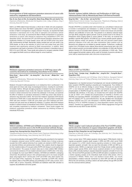
Cancer biologyB-17-159Overexpression of Nek6 suppresses premature senescence of cancer cellsinduced by camptothecin and doxorubicinHye Jin Jee, Hyun-Ju Kim, Ae Jeong Kim, Naree Song, Minjee Kim and Jeanho YunDepartment of Biochemistry, Mitochondria Hub Regulation Center, College of Medicine, Dong-AUniversity, Busan 602-714, KoreaNek6 is an NIMA-related kinase that plays a critical role in mitotic cell cycle progression.Recent studies have shown that Nek6 is upregulated in various human cancers, but thefunction of Nek6 in tumorigenesis is largely unknown. Previously, we reported that Nek6expression is decreased prior to the onset of replicative and premature cellularsenescence. In this study, we examined the effect of Nek6 overexpression on prematuresenescence of cancer cells induced by anticancer drug camptothecin (CPT) anddoxorubicin (DOX). We found that CPT and DOX-induced premature senescence weresignificantly inhibited in EJ human bladder cancer cells and H<strong>12</strong>99 human lung cancercells overexpressing HA-Nek6. Mechanistic studies revealed that cell cycle arrest in theG2/M phases, as well as the reduction of cyclin B and cdc2 protein level upon DOXtreatment were significantly reduced by Nek6 overexpression. In addition, Nek6overexpression also leads to decrease of DOX-induced increases in intracellular levels ofROS in EJ and H<strong>12</strong>99 cells. These results add evidence for oncogenic potential of Nek6and suggest that Nek6 could be an efficient target for cancer treatment.B-17-162Periostin Aptamer inhibits Adhesion and Proliferation of A549 LungAdenocarcinoma Cells by Mesenchymal Stem Cell-Derived PeriostinSang Hun Shin¹ , ², Eun Jin Seo¹and Jae Ho Kim¹ , ²¹Medical Research Center for Ischemic Tissue Regeneration, ²Department of Physiology, School ofMedicine, Pusan National University, Gyeongsangnam-do, KoreaPeriostin (POSTIN is a secreted protein which functions as a cell adhesion molecule andis involved in tumorigenesis and metastasis in a variety of cancers. Periostin is highlyexpressed in cancer-associated stromal fibroblast in human tumor tissues and promotesadhesion and proliferation of tumor cells. Thus,periostin is an attractive molecular targetand high affinity antagonists against periostin could be powerful tools for the delivery oftherapeutics and/or imaging agents to cancer patients. In this study, we screened andidentified a specific RNA aptamer, HO-2046-43-2,as a human periostin-specific aptamer.Periostin aptamer inhibits cell adhesion of A549 cells onto periostin-coated plasticdishes,but adhesion of the cells onto other extracellular matrix proteins such asfibronectin and collagen,are not inhibited by periostin aptamer. Furthermore, conditionedmedium from LPA-treated human adipose tissue-derived mesenchymal stem cells (LPACM) contained periostin and stimulated adhesion and proliferation of A549 cells.Periostinaptamer blocked LPA CM-stimulated adhesion and proliferation of A549 cells. Theseresults suggest that periostin aptamer will be useful for blockade of paracrine activation oftumor cells by mesenchymal stem cells in the tumor microenviroment.B-17-160Melatonin suppresses premature senescence of A549 lung cancer cellscaused by doxorubicin by modulating intracellular levels of ROSNaree Song¹ , ², Hyun-Ju Kim¹ , ², Ae Jeong Kim¹ , ², Hye Jin Jee¹ , ², Minjee Kim¹ , ²andJeanho Yun¹ , ²¹Department of Biochemistry, ²Mitochondria Hub Regulation Center, College of Medicine, Dong-A University, Busan 602-714, KoreaMelatonin (N-acetyl-5-methoxytryptamine) is an indoleamine that is synthesized in thepineal gland and shows wide range of physiological functions, including the coordinationof circadian rhythms. Although anti-aging properties of melatonin has been reported in asenescence-accelerated mouse model, the molecular mechanism of how melatonin isinvolved in cellular senescence has not been fully addressed. In this study, we examinedthe effect of melatonin on premature senescence of A549 lung cancer cells induced byanticancer drug doxorubicin (DOX). We found that melatonin suppresses DOX-inducedsenescence of A549 cells in a dose-dependent manner. Cell cycle analysis revealed thatA549 cells were arrested in G2/M cell cycle phase upon DOX treatment and DOXinducedcell cycle arrest was not affected by melatonin. In contrast, while DOX treatmentresulted in marked increase of intracellular levels of ROS, DOX-induced increase of ROSwas completely suppressed by melatonin cotreatment. These results suggest thatmelatonin suppresses premature cellular senescence by inhibiting increase ofintracellular ROS levels.B-17-163Effect of LHT7 on VEGFR-2Yoo-Jin Yang¹, Yoonjae Jung¹, SangMun Bae¹, Jong-Ho Kim¹, Young-Ro Byun²,Rang-Woon Park¹¹Department of Biochemistry and Cell Biology, School of Medicine, Kyungpook NationalUniversity, Daegu, Korea, and ²College of Pharmacy, Seoul National University, Seoul, KoreaAngiogenesis is an important process of tumorigenesis and vascular endothelial growthfactor receptor2 (VEGFR2) signaling is essential part of angiogenesis. Thus inhibition ofVEGFR2 can be an effective strategy to cancer therapy. Previously we reported thatLHT7, a low molecular weight heparin (LMWH) taurocholate conjugate, inhibitsphosphorylation of VEGFR2 through the interaction with VEGF in human umbilical veinendothelial cells (HUVECs). In this study, we revealed that LHT7 interact with VEGFR2as well as VEGF. LHT7 inhibited the phosphorylation of VEGFR2 in HUVECs andVEGFR2 over-expressing HEK293. Colocalization of LHT7 and VEGFR2 was observedwith FITC-labeled LHT7 by immunofluorescence. We also identified that colocalizationbetween VEGFR2 and LHT7 was decreased in VEGFR2 siRNA treated HUVECs.Binding of HT<strong>10</strong> to VEGFR2 increased in a dose-dependent manner using FACSanalysis. Therefore we suggested that LHT7 would inhibits angiogenesis through theinteract with not only VEGF but also VEGFR2.B-17-161MEK1/2 inhibitor, AS703026 and AZD6244 overcome the resistance of K-ras mutated colorectal cancer to the EGFR monoclonal antibody therapyJuyong Yoon, Kyoung-Hwa Koo, Kang-Yell ChoiTranslational Research Center for Protein Function Control, Department of Biotechnology, Collegeof Life Science and Biotechnology, Yonsei University, Seoul <strong>12</strong>0-752, KoreaA monoclonal antibodies (mAb) against EGFR are frequently prescribed for the treatmentof metastatic colorectal cancer (mCRC) patients. Multiple lines of clinical evidencesrevealed that mCRC patients harboring K-ras mutation are resistant to EGFR mAb suchas cetuximab (Erbitux) and panitumumab (Vectibix). Now, patients should be diagnosedfor their mutational status of K-ras gene before EGFR mAb therapy to avoid wasting timeand disbursement. The purpose of present study was to check whether MEK inhibitionovercomes the resistance of K-ras mutated colorectal cancer to EGFR mAb. In this study,we investigate the most recent MEK1/2 inhibitors, AS703026 and AZD6244, undergoingclinical trials. We performed various in vitro cell based assay and in vivo mouse xenograftassay using isogenic DLD-1 cell line pairs, D-WT and D-MUT cells, which express onlyWT and mutant K-Ras, respectively. As expected, cetuximab, an EGFR mAb, showedthe inhibitory effect on Ras-ERK pathway and proliferation in D-WT cells or tumors, not inD-MUT cells or tumors. Importantly, AS703026 and AZD6244 inhibit ERK activation tosuppress cell proliferation and tumor growth.B-17-164TRAIL receptor 1 and 2 induced activation of the Nox1 NADPH oxidaseand its role in the induction of caspase-independent apoptotic cell deathKyung-Jin Park, Chang-Han Lee and Yong-Sung Kim*Department of Molecular Science and Technology, Ajou University, San 5, Woncheon-dong,Yeongtong-gu, Suwon 443-749 KoreaTumor necrosis factor (TNF)-related apoptosis inducing ligand (TRAIL/Apo2L) binds itscognate pro-apoptotic receptors death receptor 4 (DR4) and DR5, which recruit FADDand caspase 8 in a DISC (death-inducing signaling complex) to initiate caspasedependentapoptotic cell death. Here we report that stimulation of DR4 and DR5 by anagonistic protein, KD548, an engineered variant from Kringle domain scaffold, generatesreactive oxygen species (ROS), which induces sustained activation of c-Jun N-terminalkinase (JNK) eventually leading to caspase-independent apoptotic cell death of tumor celllines. We found that ROS generation by DR4 and DR5 stimulation is mediated by forminga distinct signaling complex at the intracellular death domain (DD) domain, which iscomposed of TRADD, TRAF2, Nox1 and riboflavin kinase (RFK). Our resultsdemonstrates a detailed molecular mechanism by which DR4 and DR5 can generateROS at the cell membrane, providing a distinct cell death pathway through DR4 and DR5from the canonical extrinsic pathway.194 Korean Society for Biochemistry and Molecular Biology



![No 기ê´ëª
(êµë¬¸) ëíì ì íë²í¸ ì¹ì£¼ì ì·¨ê¸í목[ì문] ë¶ì¤ë²í¸ 1 ...](https://img.yumpu.com/32795694/1/190x135/no-eeeeu-e-eii-i-iei-ii-1-4-i-ieiecie-eiei-1-.jpg?quality=85)
