11:10-12:00, Rm 103
11:10-12:00, Rm 103
11:10-12:00, Rm 103
You also want an ePaper? Increase the reach of your titles
YUMPU automatically turns print PDFs into web optimized ePapers that Google loves.
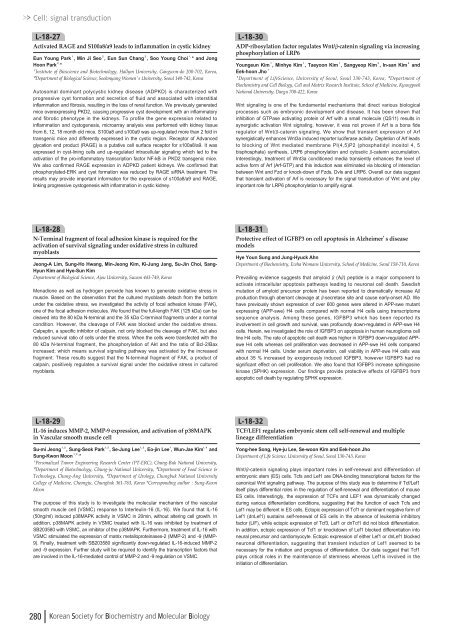
Cell: signal transductionL-18-27Activated RAGE and S<strong>10</strong>0a8/a9 leads to inflammation in cystic kidneyEun Young Park¹, Min Ji Seo², Eun Sun Chang², Soo Young Choi¹ , * and JongHoon Park² , *¹Institute of Bioscience and Biotechnology, Hallym University, Gangwon-do 2<strong>00</strong>-702, Korea,²Department of Biological Science, Sookmyung Women’s University, Seoul 140-742, KoreaAutosomal dominant polycystic kidney disease (ADPKD) is characterized withprogressive cyst formation and secretion of fluid and associated with interstitialinflammation and fibrosis, resulting in the loss of renal function. We previously generatedmice overexpressing PKD2, causing progressive cyst development with an inflammatoryand fibrotic phenotype in the kidneys. To profile the gene expression related toinflammation and cystogenesis, microarray analysis was performed with kidney tissuefrom 6, <strong>12</strong>, 18 month old mice. S<strong>10</strong>0a8 and s<strong>10</strong>0a9 was up-regulated more than 2 fold intransgenic mice and differently expressed in the cystic region. Receptor of Advancedglycation end product (RAGE) is a putative cell surface receptor for s<strong>10</strong>0a8/a9. It wasexpressed in cyst-lining cells and up-regulated intracellular signaling which led to theactivation of the pro-inflammatory transcription factor NF-kB in PKD2 transgenic mice.We also confirmed RAGE expression in ADPKD patient kidneys. We confirmed thatphosphorylated-ERK and cyst formation was reduced by RAGE siRNA treatment. Theresults may provide important information for the expression of s<strong>10</strong>0a8/a9 and RAGE,linking progressive cystogenesis with inflammation in cystic kidney.L-18-30ADP-ribosylation factor regulates Wnt/β-catenin signaling via increasingphosphorylation of LRP6Youngeun Kim¹, Minhye Kim¹, Taeyoon Kim¹, Sangyeop Kim², In-san Kim²andEek-hoon Jho¹¹Department of LifeScience, University of Seoul, Seoul 130-743, Korea, ²Department ofBiochemistry and Cell Biology, Cell and Matrix Research Institute, School of Medicine, KyungpookNational University, Daegu 7<strong>00</strong>-422, KoreaWnt signaling is one of the fundamental mechanisms that direct various biologicalprocesses such as embryonic development and disease. It has been shown thatinhibition of GTPase activating protein of Arf with a small molecule (QS<strong>11</strong>) results insynergistic activation Wnt signaling, however, it was not proven if Arf is a bona fideregulator of Wnt/β-catenin signaling. We show that transient expression of Arfsynergistically enhances Wnt3a induced reporter luciferase activity. Depletion of Arf leadsto blocking of Wnt mediated membrane PI(4,5)P2 (phosphatidyl inositol 4, 5bisphosphate) synthesis, LRP6 phosphorylation and cytosolic β-catenin accumulation.Interestingly, treatment of Wnt3a conditioned media transiently enhances the level ofactive form of Arf (Arf-GTP) and this induction was eliminated via blocking of interactionbetween Wnt and Fzd or knock-down of Fzds, Dvls and LRP6. Overall our data suggestthat transient activation of Arf is necessary for the signal transduction of Wnt and playimportant role for LRP6 phosphorylation to amplify signal.L-18-28N-Terminal fragment of focal adhesion kinase is required for theactivation of survival signaling under oxidative stress in culturedmyoblastsJeong-A Lim, Sung-Ho Hwang, Min-Jeong Kim, Ki-Jung Jang, Su-Jin Choi, Sang-Hyun Kim and Hye-Sun KimDepartment of Biological Science, Ajou University, Suwon 443-749, KoreaMenadione as well as hydrogen peroxide has known to generate oxidative stress inmuscle. Based on the observation that the cultured myoblasts detach from the bottomunder the oxidative stress, we investigated the activity of focal adhesion kinase (FAK),one of the focal adhesion molecules. We found that the full-length FAK (<strong>12</strong>5 kDa) can becleaved into the 80 kDa N-terminal and the 35 kDa C-terminal fragments under a normalcondition. However, the cleavage of FAK was blocked under the oxidative stress.Calpeptin, a specific inhibitor of calpain, not only blocked the cleavage of FAK, but alsoreduced survival ratio of cells under the stress. When the cells were transfected with the80 kDa N-terminal fragment, the phosphorylation of Akt and the ratio of Bcl-2/Baxincreased; which means survival signaling pathway was activated by the increasedfragment. These results suggest that the N-terminal fragment of FAK, a product ofcalpain, positively regulates a survival signal under the oxidative stress in culturedmyoblasts.L-18-31Protective effect of IGFBP3 on cell apoptosis in Alzheimer’s diseasemodelsHye Youn Sung and Jung-Hyuck AhnDepartment of Biochemistry, Ewha Womans University, School of Medicine, Seoul 158-7<strong>10</strong>, KoreaPrevailing evidence suggests that amyloid β(Aβ) peptide is a major component toactivate intracellular apoptosis pathways leading to neuronal cell death. Swedishmutation of amyloid precursor protein has been reported to dramatically increase Aβproduction through aberrant cleavage at β-secretase site and cause early-onset AD. Wehave previously shown expression of over 6<strong>00</strong> genes were altered in APP-swe mutantexpressing (APP-swe) H4 cells compared with normal H4 cells using transcriptomesequence analysis. Among these genes, IGFBP3 which has been reported itsinvolvement in cell growth and survival, was profoundly down-regulated in APP-swe H4cells. Herein, we investigated the role of IGFBP3 on apoptosis in human neuroglioma cellline H4 cells. The rate of apoptotic cell death was higher in IGFBP3 down-regulated APPsweH4 cells whereas cell proliferation was decreased in APP-swe H4 cells comparedwith normal H4 cells. Under serum deprivation, cell viability in APP-swe H4 cells wasabout 35 % increased by exogenously induced IGFBP3, however IGFBP3 had nosignificant effect on cell proliferation. We also found that IGFBP3 increase sphingosinekinase (SPHK) expression. Our findings provide protective effects of IGFBP3 fromapoptotic cell death by regulating SPHK expression.L-18-29IL-16 induces MMP-2, MMP-9 expression, and activation of p38MAPKin Vascular smooth muscle cellSu-mi Jeong¹ , ², Sung-Seok Park¹ , ², Se-Jung Lee¹ , ³, Eo-jin Lee¹, Wun-Jae Kim¹ , ⁴andSung-Kwon Moon¹ , ² , *¹Personalized Tumor Engineering Research Center (PT-ERC), Chung-Buk National University,²Department of Biotechnology, Chung-ju National University, ³Department of Food Science &Technology, Chung-Ang University, ⁴Department of Urology, Chungbuk National UniversityCollege of Medicine, Cheongju, Chungbuk 361-763, Korea *Corresponding author : Sung-KwonMoonThe purpose of this study is to investigate the molecular mechanism of the vascularsmooth muscle cell (VSMC) response to Interleulin-16 (IL-16). We found that IL-16(50ng/ml) induced p38MAPK activity in VSMC in 20min, without altering cell growth. Inaddition, p38MAPK activity in VSMC treated with IL-16 was inhibited by treatment ofSB203580 with VSMC, an inhibitor of the p38MAPK. Furthermore, treatment of IL-16 withVSMC stimulated the expression of matrix metalloproteinases-2 (MMP-2) and -9 (MMP-9). Finally, treatment with SB203580 significantly down-regulated IL-16-induced MMP-2and -9 expression. Further study will be required to identify the transcription factors thatare involved in the IL-16-mediated control of MMP-2 and -9 regulation on VSMC.L-18-32TCF/LEF1 regulates embryonic stem cell self-renewal and multiplelineage differentiationYong-hee Song, Hye-ju Lee, Se-woon Kim and Eek-hoon JhoDepartment of Life Science, University of Seoul, Seoul 130-743, KoreaWnt/β-catenin signaling plays important roles in self-renewal and differentiation ofembryonic stem (ES) cells. Tcfs and Lef1 are DNA-binding transcriptional factors for thecanonical Wnt signaling pathway. The purpose of this study was to determine if Tcf/Lef1itself plays differential roles in the regulation of self-renewal and differentiation of mouseES cells. Interestingly, the expression of TCFs and LEF1 was dynamically changedduring various differentiation conditions, suggesting that the function of each Tcfs andLef1 may be different in ES cells. Ectopic expression of Tcf1 or dominant negative form ofLef1 (dnLef1) sustains self-renewal of ES cells in the absence of leukemia inhibitoryfactor (LIF), while ectopic expression of Tcf3, Lef1 or dnTcf1 did not block differentiation.In addition, ectopic expression of Tcf1 or knockdown of Lef1 blocked differentiation intoneural precursor and cardiomyocyte. Ectopic expression of either Lef1 or dnLef1 blockedneuronal differentiation, suggesting that transient induction of Lef1 seemed to benecessary for the initiation and progress of differentiation. Our data suggest that Tcf1plays critical roles in the maintenance of stemness whereas Lef1is involved in theinitiation of differentiation.280 Korean Society for Biochemistry and Molecular Biology



![No 기ê´ëª
(êµë¬¸) ëíì ì íë²í¸ ì¹ì£¼ì ì·¨ê¸í목[ì문] ë¶ì¤ë²í¸ 1 ...](https://img.yumpu.com/32795694/1/190x135/no-eeeeu-e-eii-i-iei-ii-1-4-i-ieiecie-eiei-1-.jpg?quality=85)

