11:10-12:00, Rm 103
11:10-12:00, Rm 103
11:10-12:00, Rm 103
You also want an ePaper? Increase the reach of your titles
YUMPU automatically turns print PDFs into web optimized ePapers that Google loves.
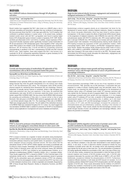
Cancer biologyB-17-<strong>12</strong>3Rsf-1 (HBXAP) induces chemoresistance through NF-κB pathwayactivationYeong-In Yang¹ , ²and Jung-Hye Choi¹ , ²¹Department of Life & Nanopharamceutical Science, Kyung Hee University, Seoul 130-701, Korea²Department of Oriental Pharmaceutical Science, College of Pharmacy, Kyung Hee University,Seoul 130-701, KoreaRecent studies have suggested that Rsf-1 (also known as a HBXAP) plays a role inchromatin remodeling and transcription regulation that may contribute to tumorigenesis.We have previously shown that Rsf-1 is the major gene within the <strong>11</strong>q13.5 amplicon thatcontributes to paclitaxel resistance in ovarian cancer. In the present study, paclitaxelresistantovarian cancer cells with Rsf-1 up-regulation were found to express higherlevels of NF-κB-dependent genes involved in evasion of apoptosis (FLIP, XIAP, BclX/L,and Bcl2) and inflammation (COX-1 and COX-2). In addition, ectopic expression of Rsf-1using tet-off inducible expression system significantly increased nuclear translocation ofNF-κB and expression of NF-κB-dependent genes. Down-regulation of Rsf-1 using shorthairpin RNA resulted in the inhibition of NF-κB-mediated anti-apoptotic gene expression.Moreover, NF-κB inhibitors Bay <strong>11</strong>70-82 and MG132 considerably enhancedchemosensitivity to paclitaxel in the Rsf-1 overexpressing OVCAR3 and Rsf-1-inducedSKOV3 cells. Taken together, these data suggest that Rsf-1 may function as acoactivator for NF-κB, consequently augmenting expression of genes necessary for thedevelopment of chemoresistance in ovarian cancer cells.B-17-<strong>12</strong>6High-fat diet-induced obesity increases angiogenesis and metastasis ofmalignant melanoma in C57BL/6 miceJae In Jung¹, You Jin Jung¹, Song Her², Jung Han Yoon Park¹¹Department of Food science and Nutrition, Hallym University, 39 Hallymdaehak-gil Chuncheon,2<strong>00</strong>-702, ²Division of Bio-Imaging, Chuncheon Center, Korea Basic Science Institute, Chuncheon2<strong>00</strong>-701, KoreaEpidemiologic evidence suggests obesity as an independent risk factor for thedevelopment of several cancers, including malignant melanoma. Obesity is associatedwith chronic low-grade inflammation which has been linked to various steps intumorigenesis. In this study to explore the effects of high-fat diet (HFD)-induced obesityon the progression of malignant melanoma, male C57BL/6N mice were fed a HFD andsubcutaneously injected with B16-F<strong>10</strong> melanoma cells. HFD caused significant weightgain in wild type C57BL/6N mice. HFD feeding did not increase solid tumor growth, butincreased lymph node metastasis. Immunofluorescence staining results indicate thatHFD feeding increased the expression of Ki67 (cell proliferation marker), CD45(macrophage marker), VEGF, VEGF receptor 2, and PECAM-1 (angiogenesis marker) intumor tissues. Western blot analysis results showed that the protein levels of COX-2,iNOS, p-Akt, p-ERK1/2, p-p38 MAPK, p-SAPK/JNK, p-STAT3, p-p65, p-cJun and HIF-1alpha were increased in the tumor tissue of mice fed on the HFD. The present resultsdemonstrate that HFD-induced obesity stimulates the inflammation, angiogenesis andmetastasis of melanoma in C57BL/6N mice.B-17-<strong>12</strong>4Growth and characterization of multicellular 3D spheroids of N2aneuroblastoma cells on RGD-incorporated elastin-like proteinKyeong-Min Lee, Mi-Ae Kwon and Won-Bae JeonLaboratory of Biochemistry and Cellular Engineering, Daegu Gyeongbuk Institute of Science andTechnology, Daegu 7<strong>11</strong>-873, KoreaMulticellular tumour spheroids (MTS) are being widely used in various aspects of tumorbiology because they mimic the growth characteristics of in vivo tumours more closelythan in vitro two-dimensional (2D) culture. The extracellular matrix (ECM) providesphysical supports for maintaining three dimensional (3D) cell morphology. However,preparation of naturally derived matrices is complicate, limited in high throughput andlarge scale production and, thus very expensive. In this study, we examined N2aneuroblastoma cells make 3D MTS on biommetic elastin-like protein consist of thetandem repeat RGD, ELP[RGD-V6]20. We have shown that ELP[RGD-V6]20 stimulatedthe spreading and proliferation of N2a cells in a manner similar to that of nativefibronectin. N2a cells grown on ELP[RGD-V6]20 started to aggregate each other after 48h, and formed spheroid shapes after 72 h. The morphology of N2a cells on ELP[RGD-V6]20 were changed grape-like structures. The cell viability of the multicellular structureson ELP[RGD-V6]20 was maintained throughout the 72 h culture. Our result suggest thatnew biomaterial, ELP[RGD-V6]20 is suitable for production of 3D MTS of neuroblastomacells for various applications in neurogenesis and cancer metastasis and treatment.B-17-<strong>12</strong>7M2-macrophages enhance tumor growth and lung metastasis ofmammary cancer cells through induction of cancer cell proliferation andmacrophage infiltrationHan Jin Cho¹, Jae In Jung¹, Song Her², Jung Han Yoon Park¹¹Department of Food Science and Nutrition, Hallym University, Chuncheon 2<strong>00</strong>-702, ²Division ofBio-Imaging, Chuncheon Center, Korea Basic Science Institute, Chuncheon 2<strong>00</strong>-701, KoreaTumor-associated macrophage (TAM) can be one of key regulators of tumormicroenvironment. TAM, generally has an M2 phenotype, is associated with poorprognosis in a variety of cancers, including breast, lung, and pancreatic cancer. In thepresent study, we examined the effect of M2-macrophages on the development ofmammary cancer using a 4T1-orthotopic tumor model. 4T1 mammary carcinoma cellswere injected into the mammary fat pad of syngeneic Balb/C mice. Co-injection of M2-macrophages increased solid tumor growth and lung metastasis. In in vivo imagingsystem using 4T1-luciferase (4T1-luc) cells, co-injection of M2-macrophages increasedbioluminescent signal in 4T1-luc tumor-bearing mice. In addition, our in vitro resultindicated that co-culture of 4T1 and M2-macrophages increased 4T1 cell proliferation.Co-culture of 4T1 cells and M2-macrophages increased the production ofchemoattractants and pro-inflammatory cytokines, resulting in the induction ofmacrophage migration. These results indicate that M2-macrophages increase tumorgrowth through induction of tumor cell proliferation and macrophage infiltration, whichleads to the promotion of metastasis.B-17-<strong>12</strong>5PTK7 is cleaved by proteases extracellularly and intracellularly andreleases an anti-angiogenic extracellular domain in colon cancer cellsHye-Won Na, Won-Sik Shin and Seung-Taek LeeDepartment of Biochemistry, College of Life Science and Biotechnology, Yonsei University, Seoul<strong>12</strong>0-749, KoreaPTK7 is known to function as a regulator of planar cell polarity during development.Although PTK7 is a kind of defective receptor protein tyrosine kinases, it enhances signaltransduction pathways induced by some growth factors. Its expression is upregulated insome malignant cancers including colorectal cancers. We have shown that theextracellular domain of PTK7 (sPTK7) inhibits migration and invasion of endothelial cellsby acting as a decoy receptor. Interestingly, we have detected that sPTK7 is secreted intoculture media of colorectal cancer cells and human embryonic kidney 293 (HEK293)cells. Generation of sPTK7 was inhibited by pan-metalloprotease inhibitors, an a-disintegrin-a-metalloprotease (ADAM)-17-selective inhibitor, and a γ-secretase inhibitor.These results suggest that PTK7 is cleaved by ADAM-17 at the C-terminal region of theextracellular domain and by γ-secretase within the transmembrane domain to generatesPTK7 into the extracellular space and cytoplasmic domain of PTK7 into cytoplasm.Since sPTK7 has the anti-angiogenic function, it is assumed that sPTK7 may have a roleto inhibit metastasis of primary tumor, similar to angiostatin and endostatin. [Supported byNRF of Korea through Basic Research, Studies on Ubiquitome Functions, and BK21programs]B-17-<strong>12</strong>8Piceatannol inhibits migration and invasion of prostate cancer cells:possible mediation by decreased interleukin-6 signalingGyoo Taik Kwon¹, Jae In Jung¹, Eun Young Woo¹, Song Her²and Jung Han YoonPark¹¹Department of Food Science and Nutrition, Hallym University, Chuncheon 2<strong>00</strong>-702, Korea,²Division of Bio-Imaging, Chuncheon Center, Korea Basic Science Institute, Chuncheon 2<strong>00</strong>-701,KoreaPiceatannol (trans-3,4,3`,5`-tetrahydroxystilbene) is a polyphenol detected in grapes, redwine, and Rheum undulatum; it has also been demonstrated to exert anticarcinogeniceffects. In this study, in order to determine whether piceatannol inhibits the lungmetastasis of prostate cancer cells, MLL rat prostate cancer cells expressing luciferasewere injected into the tail veins of male nude mice. The oral administration of piceatannol(20 mg/kg) significantly inhibited the accumulation of MLL cells in the lungs of these mice.In the cell culture studies, piceatannol was demonstrated to inhibit the basal and EGFinducedmigration and invasion of DU145 prostate cancer cells. In DU145 cells,piceatannol attenuated the secretion and mRNA levels of MMP-9, uPA and VEGF.Additionally, piceatannol inhibited the phosphorylation of STAT3. Furthermore,piceatannol effected reductions in both basal and EGF-induced IL-6 secretion. An IL-6neutralizing antibody inhibited EGF-induced STAT3 phosphorylation and EGF-stimulatedmigration of DU145 cells. These results demonstrate that the inhibition of IL-6/STAT3signaling may constitute a mechanism by which piceatannol regulates the expression ofproteins involved in regulating the migration and invasion of DU145 cells.188 Korean Society for Biochemistry and Molecular Biology



![No 기ê´ëª
(êµë¬¸) ëíì ì íë²í¸ ì¹ì£¼ì ì·¨ê¸í목[ì문] ë¶ì¤ë²í¸ 1 ...](https://img.yumpu.com/32795694/1/190x135/no-eeeeu-e-eii-i-iei-ii-1-4-i-ieiecie-eiei-1-.jpg?quality=85)


