

Chromosome segregation errors: a double-edged sword - TI Pharma
Chromosome segregation errors: a double-edged sword - TI Pharma
Chromosome segregation errors: a double-edged sword - TI Pharma

Create successful ePaper yourself
Turn your PDF publications into a flip-book with our unique Google optimized e-Paper software.
2<br />
Supplemental figures<br />
44<br />
A B<br />
C<br />
% of cells<br />
Thymidine release<br />
Thymidine release<br />
+ Mps1-IN-1<br />
% of cells with 53BP1 foci<br />
100<br />
80<br />
60<br />
40<br />
20<br />
0<br />
100<br />
80<br />
60<br />
40<br />
20<br />
0<br />
Asynchronous<br />
RO release<br />
Doxorubicin<br />
Normal nuclei<br />
Abnormal nuclei<br />
Monastrol<br />
release<br />
BJ-Tert cells<br />
>5 foci<br />
5 foci<br />
4 foci<br />
3 foci<br />
2 foci<br />
1 focus<br />
0 foci<br />
D E<br />
Mps1-IN-1<br />
(n=139)<br />
0 50 100 150 200 250 300 350<br />
Time (minutes) after anaphase<br />
% of cells<br />
100<br />
80<br />
60<br />
40<br />
20<br />
0<br />
53BP1 γ-H2AX merge w/DAPI<br />
RPE-1 cells<br />
normal<br />
% γH2AX positive cells<br />
abnormal<br />
Monastrol block<br />
100<br />
80<br />
60<br />
40<br />
20<br />
0<br />
normal<br />
abnormal<br />
1 hour 2 hours<br />
average number of 53BP1-foci/cell<br />
3,0<br />
2,5<br />
2,0<br />
1,5<br />
1,0<br />
0,5<br />
0<br />
-<br />
Control<br />
+ +<br />
Normal<br />
Abnormal<br />
Thymidine release<br />
U2OS cells<br />
Asynchronous<br />
>5 foci<br />
5 foci<br />
4 foci<br />
3 foci<br />
2 foci<br />
1 focus<br />
0 foci<br />
Mps1-IN-1<br />
Mps1-IN-1<br />
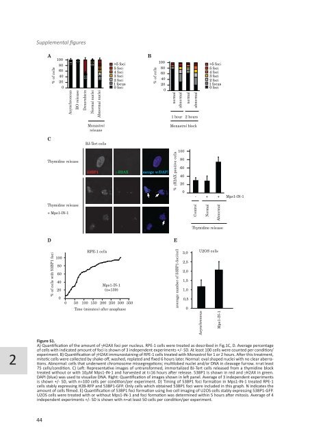
Figure S1.<br />
A) Quantification of the amount of gH2AX foci per nucleus. RPE-1 cells were treated as described in Fig.1C, D. Average percentage<br />
of cells with indicated amount of foci is shown of 3 independent experiments +/- SD. At least 100 cells were counted per condition/<br />
experiment. B) Quantification of gH2AX immunostaining of RPE-1 cells treated with Monastrol for 1 or 2 hours. After this treatment,<br />
mitotic cells were collected by shake-off, washed, replated and fixed 6 hours later. Normal: oval shaped nuclei with no clear aberrations.<br />
Abnormal: cells that underwent chromosome mis<strong>segregation</strong>s; multilobed nuclei and/or DNA in cleavage furrow. n=at least<br />
75 cells/condition. C) Left: Representative images of untransformed, immortalized BJ-Tert cells released from a thymidine block<br />
treated without or with 10mM Mps1-IN-1 and harvested at t=16 hours after release. 53BP1 is shown in red and gH2AX in green.<br />
DAPI (blue) was used to visualize DNA. Right: Quantification of images shown in left panel. Average of 3 independent experiments<br />
is shown +/- SD, with n=100 cells per condition/per experiment. D) Timing of 53BP1 foci formation in Mps1-IN-1 treated RPE-1<br />
cells stably expressing H2B-RFP and 53BP1-GFP. Only cells which obtained 53BP1 foci were included in this graph. N indicates the<br />
amount of cells filmed. E) Quantification of 53BP1 foci formation using live cell imaging of U2OS cells stably expressing 53BP1-GFP.<br />
U2OS cells were treated with or without Mps1-IN-1 and foci formation was determined within 5 hours after mitosis. Average of 4<br />
independent experiments +/- SD is shown with n=at least 50 cells per condition/per experiment.


