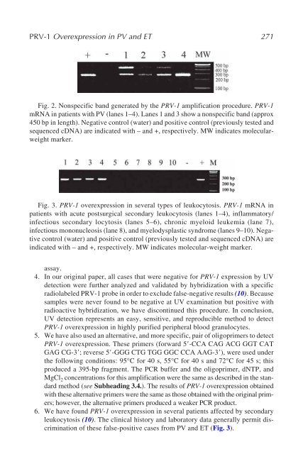
PRV-1 Overexpression in PV and ET 271 Fig. 2. Nonspecific band generated by the PRV-1 amplification procedure. PRV-1 mRNA in patients with PV (lanes 1–4). Lanes 1 and 3 show a nonspecific band (approx 450 bp in length). Negative control (water) and positive control (previously tested and sequenced cDNA) are indicated with – and +, respectively. MW indicates molecularweight marker. Fig. 3. PRV-1 overexpression in several types of leukocytosis. PRV-1 mRNA in patients with acute postsurgical secondary leukocytosis (lanes 1–4), inflammatory/ infectious secondary locytosis (lanes 5–6), chronic myeloid leukemia (lane 7), infectious mononucleosis (lane 8), and myelodysplastic syndrome (lanes 9–10). Negative control (water) and positive control (previously tested and sequenced cDNA) are indicated with – and +, respectively. MW indicates molecular-weight marker. assay. 4. In our original paper, all cases that were negative for PRV-1 expression by UV detection were further analyzed and validated by hybridization with a specific radiolabeled PRV-1 probe in order to exclude false-negative results (10). Because samples were never found to be negative at UV examination but positive with radioactive hybridization, we have discontinued this procedure. In conclusion, UV detection represents an easy, sensitive, and reproducible method to detect PRV-1 overexpression in highly purified peripheral blood granulocytes. 5. We have also used an alternative, and more specific, pair of oligoprimers to detect PRV-1 overexpression. These primers (forward 5�-CCA CAG ACG GGT CAT GAG CG-3�; reverse 5�-GGG CTG TGG GGC CCA AAG-3�), were used under the following conditions: 95°C for 40 s, 55°C for 40 s and 72°C for 45 s; this produced a 395-bp fragment. The PCR buffer and the oligoprimer, dNTP, and MgCl 2 concentrations for this amplification were the same as described in the standard method (see Subheading 3.4.). The results of PRV-1 overexpression obtained with these alternative primers were the same as those obtained with the original primers; however, the alternative primers produced a weaker PCR product. 6. We have found PRV-1 overexpression in several patients affected by secondary leukocytosis (10). The clinical history and laboratory data generally permit discrimination of these false-positive cases from PV and ET (Fig. 3).
272 Martini, Teofili, and Larocca References 1. Dameshek, W. (1951) Some speculations on the myeloproliferative syndromes. Blood 6, 372–375. 2. Adamson, J. W., Fialkow, P. J., Murphy, S., Prchal, J. F., and Steinmann, B. A. (1976) Polycythemia vera: stem-cell and probable clonal origin of the disease. N. Engl. J. Med. 295, 913–916. 3. Jacobson, R. J., Salo, A., and Fialkow, P. J. (1978) Agnogenic myeloid metaplasia: a clonal proliferation of hematopoietic stem cells with secondary myelofibrosis. Blood 51, 189–194. 4. Fialkow, P. J., Jacobson, J. R., Singer, J. W., Sacher, R. A., McGuffin, R. W., and Neefe, J. R. (1980) Philadelphia chromosome (Ph1)-negative chronic myelogenous leukemia (CML): a clonal disease with origin in a multipotent stem cell. Blood 56, 70–73. 5. Fialkow, P. J., Faguet, G. B., Jacobson, R. J., Vaidya, K., and Murphy, S. (1981) Evidence that essential thrombocythemia is a clonal disorder with origin in a multipotent stem cell. Blood 58, 916–919. 6. Berlin, N. I. (1975) Diagnosis and classification of the polycythemias. Semin. Hematol. 12, 339–351. 7. Murphy, S., Iland, H., Rosenthal, D., and Laszlo, J. (1986) Essential thrombocythemia: An interim report from the Polycythemia Vera Study Group. Semin. Hematol. 23, 177–182. 8. Temerinac, S., Klippel, S., Strunck, E., et al. (2000) Cloning of PRV-1, a novel member of the uPAR receptor superfamily, which is overexpressed in polycythemia rubra vera. Blood 95, 2569–2576. 9. Klippel, S., Strunck, E., Busse, C.E., Behringer, D. and Pahl, H.L. (2002) Biochemical characterization of PRV-1, a novel hematopoietic cell surface receptor, which is overexpressed in polycythemia rubra vera. Blood 100, 2441- 2448. 10. Teofili, L., Martini, M., Luongo, M., et al. (2002) Overexpression of the Polycythemia Rubra Vera-1 Gene in Essential Thrombocythemia. J. Clin. Oncol. 20, 4249–4254. 11. Johansson, P., Andreasson, B., Safai-Kutti, S., et al. (2003) The presence of a significant association between elevated PRV-1 mRNA expression and low plasma erythropoietin concentration in essential thrombocythaemia. Eur. J. Haematol. 70, 358–362. 12. Liu, E., Jelinek, J., Pastore, Y. D., Guan, Y., Prchal, J. F., and Prchal, J. T. (2003) Discrimination of polycythemias and thrombocytoses by novel, simple, accurate clonality assays and comparison with PRV-1 expression and BFU-E response to erythropoietin. Blood 101, 3294–3301. 13. Kralovics, R., Buser, A. S., Teo, S. S., et al. (2003) Comparison of molecular markers in a cohort of patients with chronic myeloproliferative disorders. Blood 102, 1869–1871. 14. Klippel, S., Strunck, E., Temerinac, S., et al. (2003) Quantification of PRV-1 mRNA distinguishes polycythemia vera from secondary erythrocytosis. Blood 102, 3569–3574.
- Page 1 and 2:
M E T H O D S I N M O L E C U L A R
- Page 3 and 4:
M E T H O D S I N M O L E C U L A R
- Page 5 and 6:
© 2006 Humana Press Inc. 999 River
- Page 7 and 8:
vi Preface comprehensive review of
- Page 9 and 10:
viii Contents 11 Detection of the F
- Page 11 and 12:
x Contributors D. GARY GILLILAND
- Page 14 and 15:
RNA and DNA From Leukocytes 1 1 Iso
- Page 16 and 17:
RNA and DNA From Leukocytes 3 mine
- Page 18 and 19:
RNA and DNA From Leukocytes 5 late
- Page 20 and 21:
RNA and DNA From Leukocytes 7 9. Us
- Page 22 and 23:
RNA and DNA From Leukocytes 9 16. C
- Page 24 and 25:
RNA and DNA From Leukocytes 11 3. I
- Page 26 and 27:
Cytogenetic and FISH Techniques 13
- Page 28 and 29:
Cytogenetic and FISH Techniques 15
- Page 30 and 31:
Cytogenetic and FISH Techniques 17
- Page 32 and 33:
Cytogenetic and FISH Techniques 19
- Page 34 and 35:
Cytogenetic and FISH Techniques 21
- Page 36 and 37:
Cytogenetic and FISH Techniques 23
- Page 38 and 39:
Cytogenetic and FISH Techniques 25
- Page 40 and 41:
Real-Time RT-PCR Strategies 27 3 Ov
- Page 42 and 43:
Real-Time RT-PCR Strategies 29
- Page 44 and 45:
Real-Time RT-PCR Strategies 31 cifi
- Page 46 and 47:
Real-Time RT-PCR Strategies 33 2.1.
- Page 48 and 49:
Real-Time RT-PCR Strategies 35 Fig.
- Page 50 and 51:
Real-Time RT-PCR Strategies 37 2.1.
- Page 52 and 53:
Real-Time RT-PCR Strategies 39 the
- Page 54 and 55:
Real-Time RT-PCR Strategies 41 Fig.
- Page 56 and 57:
Real-Time RT-PCR Strategies 43 duri
- Page 58 and 59:
MyiQ single color 400 nm 585 nm 96
- Page 60 and 61:
Real-Time RT-PCR Strategies 47 side
- Page 62 and 63:
Table 2 Real-Time Quantitative Poly
- Page 64 and 65:
Real-Time RT-PCR Strategies 51 AML-
- Page 66 and 67:
Real-Time RT-PCR Strategies 53 the
- Page 68 and 69:
Real-Time RT-PCR Strategies 55 plas
- Page 70 and 71:
Real-Time RT-PCR Strategies 57 betw
- Page 72 and 73:
Real-Time RT-PCR Strategies 59 Ct C
- Page 74 and 75:
Real-Time RT-PCR Strategies 61 Fig.
- Page 76 and 77:
Real-Time RT-PCR Strategies 63 asse
- Page 78 and 79:
Real-Time RT-PCR Strategies 65 21.
- Page 80 and 81:
Real-Time RT-PCR Strategies 67 50.
- Page 82 and 83:
Quantitative PCR for Monitoring CML
- Page 84 and 85:
Quantitative PCR for Monitoring CML
- Page 86 and 87:
Quantitative PCR for Monitoring CML
- Page 88 and 89:
Quantitative PCR for Monitoring CML
- Page 90 and 91:
Quantitative PCR for Monitoring CML
- Page 92 and 93:
Quantitative PCR for Monitoring CML
- Page 94 and 95:
Quantitative PCR for Monitoring CML
- Page 96 and 97:
Quantitative PCR for Monitoring CML
- Page 98 and 99:
Quantitative PCR for Monitoring CML
- Page 100 and 101:
Quantitative PCR for Monitoring CML
- Page 102 and 103:
Quantitative PCR for Monitoring CML
- Page 104 and 105:
Quantitative PCR for Monitoring CML
- Page 106 and 107:
Detection of BCR-ABL Mutations 93 5
- Page 108 and 109:
Detection of BCR-ABL Mutations 95 F
- Page 110 and 111:
Detection of BCR-ABL Mutations 97 2
- Page 112 and 113:
Detection of BCR-ABL Mutations 99 o
- Page 114 and 115:
Detection of BCR-ABL Mutations 101
- Page 116 and 117:
Detection of BCR-ABL Mutations 103
- Page 118 and 119:
Detection of BCR-ABL Mutations 105
- Page 120 and 121:
Deletion of Derivative Chromosome 9
- Page 122 and 123:
Deletion of Derivative Chromosome 9
- Page 124 and 125:
Deletion of Derivative Chromosome 9
- Page 126 and 127:
Deletion of Derivative Chromosome 9
- Page 128 and 129:
Diagnosis of PML-RARA-Positive APL
- Page 130 and 131:
Diagnosis of PML-RARA-Positive APL
- Page 132 and 133:
Diagnosis of PML-RARA-Positive APL
- Page 134 and 135:
Diagnosis of PML-RARA-Positive APL
- Page 136 and 137:
Diagnosis of PML-RARA-Positive APL
- Page 138 and 139:
Diagnosis of PML-RARA-Positive APL
- Page 140 and 141:
Duplexed QZyme RT-PCR for APL Analy
- Page 142 and 143:
Duplexed QZyme RT-PCR for APL Analy
- Page 144 and 145:
Duplexed QZyme RT-PCR for APL Analy
- Page 146 and 147:
Duplexed QZyme RT-PCR for APL Analy
- Page 148 and 149:
Duplexed QZyme RT-PCR for APL Analy
- Page 150 and 151:
Duplexed QZyme RT-PCR for APL Analy
- Page 152 and 153:
Duplexed QZyme RT-PCR for APL Analy
- Page 154 and 155:
Duplexed QZyme RT-PCR for APL Analy
- Page 156 and 157:
Duplexed QZyme RT-PCR for APL Analy
- Page 158 and 159:
Duplexed QZyme RT-PCR for APL Analy
- Page 160 and 161:
Duplexed QZyme RT-PCR for APL Analy
- Page 162 and 163:
Diagnosis of AML1-MTG8 (ETO)-Positi
- Page 164 and 165:
Diagnosis of AML1-MTG8 (ETO)-Positi
- Page 166 and 167:
Diagnosis of AML1-MTG8 (ETO)-Positi
- Page 168 and 169:
Diagnosis of AML1-MTG8 (ETO)-Positi
- Page 170 and 171:
Diagnosis of AML1-MTG8 (ETO)-Positi
- Page 172 and 173:
Diagnosis of AML1-MTG8 (ETO)-Positi
- Page 174 and 175:
Diagnosis of AML1-MTG8 (ETO)-Positi
- Page 176 and 177:
Diagnosis of CBFB-MYH11-Positive AM
- Page 178 and 179:
165 Table 1 Primers for CBFB-MYH11
- Page 180 and 181:
Diagnosis of CBFB-MYH11-Positive AM
- Page 182 and 183:
Diagnosis of CBFB-MYH11-Positive AM
- Page 184 and 185:
Diagnosis of CBFB-MYH11-Positive AM
- Page 186 and 187:
Diagnosis of CBFB-MYH11-Positive AM
- Page 188 and 189:
Diagnosis of CBFB-MYH11-Positive AM
- Page 190 and 191:
FIP1L1-PDGFRA in Hypereosinophilic
- Page 192 and 193:
FIP1L1-PDGFRA in Hypereosinophilic
- Page 194 and 195:
FIP1L1-PDGFRA in Hypereosinophilic
- Page 196 and 197:
FIP1L1-PDGFRA in Hypereosinophilic
- Page 198 and 199:
FIP1L1-PDGFRA in Hypereosinophilic
- Page 200 and 201:
FIP1L1-PDGFRA in Hypereosinophilic
- Page 202 and 203:
FLT3 Mutations in AML 189 12 FLT3 M
- Page 204 and 205:
FLT3 Mutations in AML 191 3.1.1. DN
- Page 206 and 207:
FLT3 Mutations in AML 193 Table 2 R
- Page 208 and 209:
FLT3 Mutations in AML 195 Fig. 2. D
- Page 210 and 211:
FLT3 Mutations in AML 197 10. Kiyoi
- Page 212 and 213:
WT1 Expression in AML and MDS 199 1
- Page 214 and 215:
WT1 Expression in AML and MDS 201 T
- Page 216 and 217:
WT1 Expression in AML and MDS 203 1
- Page 218 and 219:
WT1 Expression in AML and MDS 205 T
- Page 220 and 221:
WT1 Expression in AML and MDS 207 F
- Page 222 and 223:
WT1 Expression in AML and MDS 209 2
- Page 224 and 225:
WT1 Expression in AML and MDS 211 1
- Page 226 and 227:
Classification of AML by DNA-Oligon
- Page 228 and 229:
Classification of AML by DNA-Oligon
- Page 230 and 231:
Classification of AML by DNA-Oligon
- Page 232 and 233:
Classification of AML by DNA-Oligon
- Page 234 and 235: Classification of AML by DNA-Oligon
- Page 236 and 237: Classification of AML by DNA-Oligon
- Page 238 and 239: Classification of AML by DNA-Oligon
- Page 240 and 241: Classification of AML by DNA-Oligon
- Page 242 and 243: Classification of AML by DNA-Oligon
- Page 244 and 245: Classification of AML by DNA-Oligon
- Page 246 and 247: 233 Classification of AML by DNA-Ol
- Page 248 and 249: Classification of AML by DNA-Oligon
- Page 250 and 251: Classification of AML by DNA-Oligon
- Page 252 and 253: Classification of AML by DNA-Oligon
- Page 254 and 255: Classification of AML by Monoclonal
- Page 256 and 257: Classification of AML by Monoclonal
- Page 258 and 259: Classification of AML by Monoclonal
- Page 260 and 261: 247 Classification of AML by Monocl
- Page 262 and 263: Classification of AML by Monoclonal
- Page 264 and 265: Classification of AML by Monoclonal
- Page 266 and 267: Detecting JAK2 V617F Mutation in MP
- Page 268 and 269: Detecting JAK2 V617F Mutation in MP
- Page 270 and 271: Detecting JAK2 V617F Mutation in MP
- Page 272 and 273: Detecting JAK2 V617F Mutation in MP
- Page 274 and 275: Detecting JAK2 V617F Mutation in MP
- Page 276 and 277: Detecting JAK2 V617F Mutation in MP
- Page 278 and 279: PRV-1 Overexpression in PV and ET 2
- Page 280 and 281: PRV-1 Overexpression in PV and ET 2
- Page 282 and 283: PRV-1 Overexpression in PV and ET 2
- Page 286 and 287: PRV-1 Overexpression in PV and ET 2
- Page 288 and 289: Chimerism Analysis 275 18 Chimerism
- Page 290 and 291: Chimerism Analysis 277 1.2. Detecti
- Page 292 and 293: Chimerism Analysis 279 3. Extractio
- Page 294 and 295: Chimerism Analysis 281 14. 310 Gene
- Page 296 and 297: Chimerism Analysis 283 12. Upon com
- Page 298 and 299: Chimerism Analysis 285 Fig. 2. Calc
- Page 300 and 301: Chimerism Analysis 287 4. Allow the
- Page 302 and 303: Chimerism Analysis 289 capillary el
- Page 304 and 305: Chimerism Analysis 291 and recipien
- Page 306 and 307: Chimerism Analysis 293 Table 2 Comm
- Page 308: Chimerism Analysis 295 12. Maas, F.
- Page 311 and 312: 298 Index management, 128 AML, see
- Page 313 and 314: 300 Index and AML, 213, 236, 238, 2
- Page 315 and 316: 302 Index chimerism analysis in sex
- Page 317 and 318: 304 Index minimal residual disease
- Page 319: 306 Index Stem cell transplantation