


Create successful ePaper yourself
Turn your PDF publications into a flip-book with our unique Google optimized e-Paper software.
30 Picard, Silvy, and Gabert<br />
2. Principles of RQ-PCR<br />
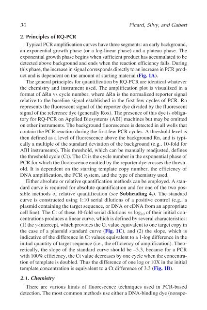
Typical PCR amplification curves have three segments: an early background,<br />
an exponential growth phase (or a log-linear phase) and a plateau phase. The<br />
exponential growth phase begins when sufficient product has accumulated to be<br />
detected above background and ends when the reaction efficiency falls. During<br />
this phase, the increase in signal corresponds directly to an increase in PCR product<br />
and is dependent on the amount of starting material (Fig. 1A).<br />
The general principles for quantification by RQ-PCR are identical whatever<br />
the chemistry and instrument used. The amplification plot is visualized in a<br />
format of ∆Rn vs cycle number, where ∆Rn is the normalized reporter signal<br />
relative to the baseline signal established in the first few cycles of PCR. Rn<br />
represents the fluorescent signal of the reporter dye divided by the fluorescent<br />
signal of the reference dye (generally Rox). The presence of this dye is obligatory<br />
for RQ-PCR on Applied Biosystems (ABI) machines but may be omitted<br />
on other instruments. The background fluorescence is detected in all wells that<br />
contain the PCR reaction during the first few PCR cycles. A threshold level is<br />
then defined as a level of fluorescence above the background Rn, and is typically<br />
a multiple of the standard deviation of the background (e.g., 10-fold for<br />
ABI instruments). This threshold, which can be manually readjusted, defines<br />
the threshold cycle (Ct). The Ct is the cycle number in the exponential phase of<br />
PCR for which the fluorescence emitted by the reporter dye crosses the threshold.<br />
It is dependent on the starting template copy number, the efficiency of<br />
DNA amplification, the PCR system, and the type of chemistry used.<br />
Either absolute or relative quantification methods can be employed. A standard<br />
curve is required for absolute quantification and for one of the two possible<br />
methods of relative quantification (see Subheading 4.). The standard<br />
curve is constructed using 1:10 serial dilutions of a positive control (e.g., a<br />
plasmid containing the target sequence, or DNA or cDNA from an appropriate<br />
cell line). The Ct of these 10-fold serial dilutions vs log 10 of their initial concentrations<br />
produces a linear curve, which is defined by several characteristics:<br />
(1) the y-intercept, which provides the Ct value equivalent to one target copy in<br />
the case of a plasmid standard curve (Fig. 1C), and (2) the slope, which is<br />
indicative of the difference in Ct values equivalent to a 1-log difference in the<br />
initial quantity of target sequence (i.e., the efficiency of amplification). Theoretically,<br />
the slope of the standard curve should be –3.3, because for a PCR<br />
with 100% efficiency, the Ct value decreases by one cycle when the concentration<br />
of template is doubled. Thus the difference of one log or 10X in the initial<br />
template concentration is equivalent to a Ct difference of 3.3 (Fig. 1B).<br />
2.1. Chemistry<br />
There are various kinds of fluorescence techniques used in PCR-based<br />
detection. The most common methods use either a DNA-binding dye (nonspe-


