MIOLO DO HUGV 2007.pmd - Hospital Universitário Getúlio Vargas
MIOLO DO HUGV 2007.pmd - Hospital Universitário Getúlio Vargas
MIOLO DO HUGV 2007.pmd - Hospital Universitário Getúlio Vargas
- No tags were found...

You also want an ePaper? Increase the reach of your titles
YUMPU automatically turns print PDFs into web optimized ePapers that Google loves.
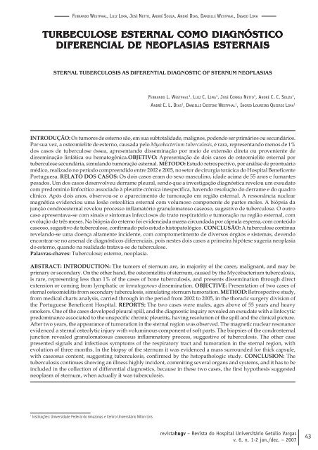
FERNAN<strong>DO</strong> WESTPHAL, LUIZ LIMA, JOSÉ NETTO, ANDRÉ SOUZA, ANDRÉ DIAS, DANIELLE WESTPHAL, INGRID LIMATURBECULOSE ESTERNTERNAL COMO DIAGNGNÓSÓSTICTICODIFERENCIAL DE NEOPLASIAS ESTERNTERNAISSTERNAL TUBERCULOSIS AS DIFERENTIAL DIAGNOSTIC OF STERNUM NEOPLASIASFERNAN<strong>DO</strong> L. WESTPHAL 1 , LUIZ C. LIMA 1 , JOSÉ CORREA NETTO 1 , ANDRÉ C. C. SOUZA 1 ,ANDRÉ C. L. DIAS 1 , DANIELLE CRISTINE WESTPHAL 1 , INGRID LOUREIRO QUEIROZ LIMA 1INTRODUÇÃO: Os tumores de esterno são, em sua subtotalidade, malignos, podendo ser primários ou secundários.Por sua vez, a osteomielite de esterno, causada pelo Mycobacterium tuberculosis, é rara, representando menos de 1%dos casos de tuberculose óssea, apresentando disseminação por meio de extensão direta ou proveniente dedisseminação linfática ou hematogênica.OBJETIVO: Apresentação de dois casos de osteomielite esternal portuberculose secundária, simulando tumoração esternal. MÉTO<strong>DO</strong>: Estudo retrospectivo, por análise de prontuáriomédico, realizado no período compreendido entre 2002 e 2005, no setor de cirurgia torácica do <strong>Hospital</strong> BeneficentePortuguesa. RELATO <strong>DO</strong>S CASOS: Os dois casos eram do sexo masculino, idade acima de 55 anos e fumantespesados. Um dos casos desenvolveu derrame pleural, sendo que a investigação diagnóstica revelou um exsudatocom predomínio linfocítico associado à pleurite crônica inespecífica, havendo resolução do derrame e do quadroclínico. Após dois anos, observou-se o aparecimento de tumoração em região esternal. A ressonância nuclearmagnética evidenciou uma lesão osteolítica esternal com volumoso componente de partes moles. A biópsia dajunção condroesternal revelou processo inflamatório granulomatoso caseoso, sugestivo de tuberculose. O outrocaso apresentava-se com sinais e sintomas infecciosos do trato respiratório e tumoração na região esternal, comevolução de três meses. Na biópsia do esterno foi evidenciada massa circundada por cápsula espessa, com conteúdocaseoso, sugestivo de tuberculose, confirmado pelo estudo histopatológico. CONCLUSÃO: A tuberculose continuarevelando-se uma doença altamente incidente, com comprometimento de diversos órgãos e sistemas, devendoencontrar-se no arsenal de diagnósticos diferenciais, pois nestes dois casos a primeira hipótese sugeria neoplasiado esterno, quando na realidade tratava-se de tuberculose.Palavras-chaves: Tuberculose; esterno, neoplasia.ABSTRACT: INTRODUCTION: The tumors of sternum are, in majority of the cases, malignant, and may beprimary or secondary. On the other hand, the osteomielitis of sternum, caused by the Mycobacterium tuberculosis,is rare, representing less than 1% of the cases of bone tuberculosis, and presents dissemination through directextension or coming from lymphatic or hematogenous dissemination. OBJECTIVE: Presentation of two cases ofsternal osteomielitis from secondary tuberculosis, simulating sternum tumoration. METHOD: Retrospective study,from medical charts analysis, carried through in the period from 2002 to 2005, in the thoracic surgery division ofthe Portuguese Beneficent <strong>Hospital</strong>. REPORTS: The two cases were males, ages above of 55 years and heavysmokers. One of the cases developed pleural spill, and the diagnostic inquiry revealed an exsudate with a linfocyticpredominance associated to the unspecific chronic pleuritis, having resolution of the spill and the clinical picture.After two years, the appearance of tumoration in the sternal region was observed. The magnetic nuclear resonanceevidenced a sternal osteolytic injury with voluminous component of soft parts. The biopsies of the condrosternaljunction revealed granulomatosus caseosus inflammatory process, suggestive of tuberculosis. The other casepresented signals and infectious symptoms of the respiratory tract and tumoration in the sternal region, withevolution of three months. In the biopsy of the sternum it was evidenced a mass surrounded for thick capsule,with caseosus content, suggesting tuberculosis, confirmed by the hstopathologic study. CONCLUSION: Thetuberculosis continues showing an illness highly incident, commiting several organs and systems, and it has to beincluded in the collection of differential diagnostics, because in these two cases, the first hypothesis suggestedneoplasm of sternum, when actually it was tuberculosis.1Instituições: Universidade Federal do Amazonas e Centro Universitário Nilton Linsrevistahugv – Revista do <strong>Hospital</strong> Universitário Getúlio <strong>Vargas</strong>v. 6. n. 1-2 jan./dez. – 200743