OM t of c.iii - Vision Research Coordinating Center - Washington ...
OM t of c.iii - Vision Research Coordinating Center - Washington ...
OM t of c.iii - Vision Research Coordinating Center - Washington ...
Create successful ePaper yourself
Turn your PDF publications into a flip-book with our unique Google optimized e-Paper software.
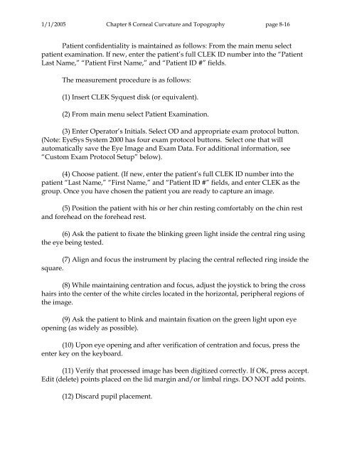
1/1/2005 Chapter 8 Corneal Curvature and Topography page 8-16<br />
Patient confidentiality is maintained as follows: From the main menu select<br />
patient examination. If new, enter the patient’s full CLEK ID number into the “Patient<br />
Last Name,” “Patient First Name,” and “Patient ID #” fields.<br />
The measurement procedure is as follows:<br />
(1) Insert CLEK Syquest disk (or equivalent).<br />
(2) From main menu select Patient Examination.<br />
(3) Enter Operator’s Initials. Select OD and appropriate exam protocol button.<br />
(Note: EyeSys System 2000 has four exam protocol buttons. Select one that will<br />
automatically save the Eye Image and Exam Data. For additional information, see<br />
“Custom Exam Protocol Setup” below).<br />
(4) Choose patient. (If new, enter the patient’s full CLEK ID number into the<br />
patient “Last Name,” “First Name,” and “Patient ID #” fields, and enter CLEK as the<br />
group. Once you have chosen the patient you are ready to capture an image.<br />
(5) Position the patient with his or her chin resting comfortably on the chin rest<br />
and forehead on the forehead rest.<br />
(6) Ask the patient to fixate the blinking green light inside the central ring using<br />
the eye being tested.<br />
(7) Align and focus the instrument by placing the central reflected ring inside the<br />
square.<br />
(8) While maintaining centration and focus, adjust the joystick to bring the cross<br />
hairs into the center <strong>of</strong> the white circles located in the horizontal, peripheral regions <strong>of</strong><br />
the image.<br />
(9) Ask the patient to blink and maintain fixation on the green light upon eye<br />
opening (as widely as possible).<br />
(10) Upon eye opening and after verification <strong>of</strong> centration and focus, press the<br />
enter key on the keyboard.<br />
(11) Verify that processed image has been digitized correctly. If OK, press accept.<br />
Edit (delete) points placed on the lid margin and/or limbal rings. DO NOT add points.<br />
(12) Discard pupil placement.


