

OM t of c.iii - Vision Research Coordinating Center - Washington ...
OM t of c.iii - Vision Research Coordinating Center - Washington ...
OM t of c.iii - Vision Research Coordinating Center - Washington ...

Create successful ePaper yourself
Turn your PDF publications into a flip-book with our unique Google optimized e-Paper software.
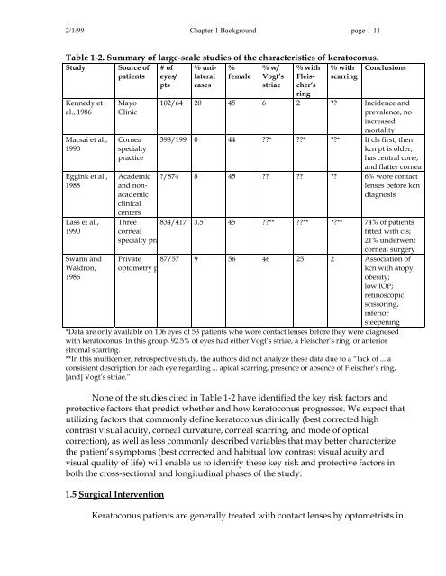
2/1/99 Chapter 1 Background page 1-11<br />
Table 1-2. Summary <strong>of</strong> large-scale studies <strong>of</strong> the characteristics <strong>of</strong> keratoconus.<br />
Study Source <strong>of</strong><br />
patients<br />
%<br />
female<br />
% with<br />
scarring<br />
Conclusions<br />
Kennedy et<br />
al., 1986<br />
Macsai et al.,<br />
1990<br />
Eggink et al.,<br />
1988<br />
Lass et al.,<br />
1990<br />
Swann and<br />
Waldron,<br />
1986<br />
Mayo<br />
Clinic<br />
Cornea<br />
specialty<br />
practice<br />
Academic<br />
and nonacademic<br />
clinical<br />
centers<br />
# <strong>of</strong><br />
eyes/<br />
pts<br />
% unilateral<br />
cases<br />
% w/<br />
Vogt’s<br />
striae<br />
% with<br />
Fleischer’s<br />
ring<br />
102/64 20 45 6 2 ?? Incidence and<br />
prevalence, no<br />
increased<br />
mortality<br />
398/199 0 44 ??* ??* ??* If cls first, then<br />
kcn pt is older,<br />
has central cone,<br />
and flatter cornea<br />
?/874 8 45 ?? ?? ?? 6% wore contact<br />
lenses before kcn<br />
diagnosis<br />
Three<br />
corneal<br />
specialty pra<br />
834/417 3.5 45 ??** ??** ??** 74% <strong>of</strong> patients<br />
fitted with cls;<br />
21% underwent<br />
corneal surgery<br />
Private<br />
optometry p<br />
87/57 9 56 46 25 2 Association <strong>of</strong><br />
kcn with atopy,<br />
obesity;<br />
low IOP;<br />
retinoscopic<br />
scissoring,<br />
inferior<br />
steepening<br />
*Data are only available on 106 eyes <strong>of</strong> 53 patients who wore contact lenses before they were diagnosed<br />
with keratoconus. In this group, 92.5% <strong>of</strong> eyes had either Vogt’s striae, a Fleischer’s ring, or anterior<br />
stromal scarring.<br />
**In this multicenter, retrospective study, the authors did not analyze these data due to a “lack <strong>of</strong> ... a<br />
consistent description for each eye regarding ... apical scarring, presence or absence <strong>of</strong> Fleischer’s ring,<br />
[and] Vogt’s striae.”<br />
None <strong>of</strong> the studies cited in Table 1-2 have identified the key risk factors and<br />
protective factors that predict whether and how keratoconus progresses. We expect that<br />
utilizing factors that commonly define keratoconus clinically (best corrected high<br />
contrast visual acuity, corneal curvature, corneal scarring, and mode <strong>of</strong> optical<br />
correction), as well as less commonly described variables that may better characterize<br />
the patient’s symptoms (best corrected and habitual low contrast visual acuity and<br />
visual quality <strong>of</strong> life) will enable us to identify these key risk and protective factors in<br />
both the cross-sectional and longitudinal phases <strong>of</strong> the study.<br />
1.5 Surgical Intervention<br />
Keratoconus patients are generally treated with contact lenses by optometrists in


