


You also want an ePaper? Increase the reach of your titles
YUMPU automatically turns print PDFs into web optimized ePapers that Google loves.
64<br />
<strong>My</strong> PET scan results<br />
Friday, April 22, 2011<br />
I went for my PET scan today. I have been having increasing discomfort in my lower<br />
chest and anticipated that my posterior mediastinal mass would be larger and pressing<br />
on my distal esophagus causing the symptoms. I was hoping there would be no evidence<br />
of new disease. This <strong>Merkel</strong> <strong>Cell</strong> carcinoma can grow as fast as any malignancy I know<br />
about.<br />
I had to be on a sugar-free diet for 24 hours prior to the examination. Last night, we had<br />
Gary, Dana, Eva, Sara and Bel over for a mini-Seder. I could not enjoy much of the<br />
cooking, but everyone had a good time.<br />
No breakfast this morning. I arrived for my scans at 8:15. They put me in a very cold<br />
room <strong>with</strong> a heating pad on my arm to start the IV. Of course, in the cold, your veins<br />
constrict, but the IV was started <strong>with</strong>out incident. <strong>My</strong> blood sugar was 105, a good<br />
number. They injected the radioisotope <strong>with</strong>out incident and then I had to wait 1 hour for<br />
it to spread through my body. It concentrates in areas of high metabolic activity, such as<br />
tumors, healing surgery sites, brain and kidneys. There was alot of activity in my brain,<br />
probably, like Cassius from Julius Caesar, I think too much.<br />
After the hour, it is 35 minutes to be scanned <strong>with</strong> the warning to stay perfectly still to<br />
avoid image degradation. Of course, as soon as the scans began, my ear and my nose<br />
began to itch. I needed to clear my throat, and the blowing of cool air over me for<br />
ventilation made my face itchy and gave me the urge to sneeze. 35 minutes seemed like<br />
hours. I left at about 11:30 AM and returned at 2:00 PM to review the results <strong>with</strong> Dr. Dan<br />
Stobbe. He is a longtime colleague and friend.<br />
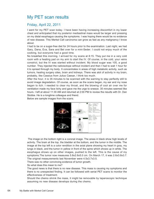
Below are sample images from the scans.<br />
The image on the bottom right is a coronal image. The areas in black show high levels of<br />
activity. The brain at the top, the bladder at the bottom, and the tumor in the middle. The<br />
image at the top left is a color rendition in the axial plane showing my heart in gray, my<br />
lungs in black, and the tumor in yellow in front of the spine which shows up in white. The<br />
esophagus shows up on other images, pushed to the left. This is the cause of my<br />
symptoms.The tumor now measures 5.8x2.6x5.0 cm. On March 17, it was 2.6x3.8x3.7.<br />
The original measurements last November were 4.5x3.7x3.5.<br />
There was no other convincing evidence of tumor growth.<br />
So what does this mean to me?<br />
The good news is that there is no new disease. This mass is causing my symptoms and<br />
there is no unexpected finding. It can be followed <strong>with</strong> serial PET scans to monitor the<br />
effectiveness of treatment.<br />
Should the chemo shrink the mass, it might be removable by laparoscopic technique<br />
assuming no new disease develops during the chemo.<br />
<strong>My</strong> <strong>Battle</strong> <strong>with</strong> <strong>Merkel</strong> <strong>Cell</strong> <strong>Cancer</strong>