The Genom of Homo sapiens.pdf
The Genom of Homo sapiens.pdf
The Genom of Homo sapiens.pdf
- TAGS
- homo
- www.yumpu.com

Create successful ePaper yourself
Turn your PDF publications into a flip-book with our unique Google optimized e-Paper software.
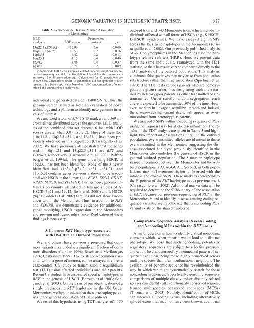
GENOMIC VARIATION IN MULTIGENIC TRAITS: HSCR 377Table 2. <strong>Genom</strong>e-wide Disease Marker Associationin MennonitesMLDProportionanalysis LOD score mutant p13q22.3 (EDNRB) 110.96 0.6 0.00010q11.21 (RET) 18.53 0.2 0.01611p15.3 6.42 0.2 0.01116q23.1 4.15 0.4 0.0461p34.3 3.86 0.4 0.0374q31.1 3.71 0.2 0.009<strong>Genom</strong>e-wide LOD scores were calculated under assumptions that locusheterogeneity was 0.2, 0.4, 0.6, 0.8, or 1.0 and that the disease variantarose 12 or 48 generations ago. Calculations for 12 generations areshown here. Calculations under 48 generations did not appreciably alterresults. p is a bootstrap p value based on 1,000 randomizations <strong>of</strong> transmittedand untransmitted haplotypes.individual and generated data on ~1,400 SNPs. Thus, thegenome screen served as both an evaluation <strong>of</strong> noveltechnology and a platform to identify new genomic intervals<strong>of</strong> interest.We analyzed a total <strong>of</strong> 5,747 SNP markers and 569 microsatellitesdistributed across the genome. MLD analysis<strong>of</strong> the combined data set detected 6 loci with LODscores greater than 3.0 (Table 2). Three <strong>of</strong> these loci(10q11.21, 13q22.3-q31.1, and 16q23.1) have been previouslyobserved in this population (Carrasquillo et al.2002). We have previously demonstrated that the geneswithin 10q11.21 and 13q22.3-q31.1 are RET andEDNRB, respectively (Carrasquillo et al. 2002, Puffenbergeret al. 1994a). <strong>The</strong> gene underlying HSCR in16q23.1 has not been identified. None <strong>of</strong> the 3 newlyidentified loci (1p34.3-p34.2, 4q31.1-q31.21, and11p15.3) contains genes previously shown to be associatedwith HSCR in the human (i.e., ECE1, EDN3, GDNF,NRTN, SOX10, and ZFHX1B). Furthermore, genomic intervalspreviously identified in linkage studies <strong>of</strong> S-HSCR (3p21 and 19q12; Bolk et al. 2000) and L-HSCR(9q31; Gabriel et al. 2002) families did not show associationwithin the Mennonites. Thus, in addition to RETand EDNRB, we demonstrate evidence for additionalgenes modifying HSCR expression in the Mennonitesand proving multigenic inheritance. Replication <strong>of</strong> thesefindings is necessary.A Common RET Haplotype Associatedwith HSCR in an Outbred PopulationWe, and others, have previously proposed that commonvariants may underlie a significant fraction <strong>of</strong> commondisorders (Lander 1996; Risch and Merikangas1996; Chakravarti 1999). <strong>The</strong> existence <strong>of</strong> common variants,within a gene <strong>of</strong> interest, can be assayed in either acase-control (CS) study or transmission disequilibriumtest (TDT) using affected individuals and their parents.Recent CS studies have associated specific haplotypes inRET in the genesis <strong>of</strong> HSCR (Borrego et al. 2003; Sancandiet al. 2003). On the basis <strong>of</strong> our identification <strong>of</strong> asingle predisposing RET haplotype in the Old OrderMennonites, we hypothesized that the same haplotype existsin the general population <strong>of</strong> HSCR patients.We tested this hypothesis using TDT analyses <strong>of</strong> >150outbred trios and ~43 Mennonite trios, which include individualsaffected with all forms <strong>of</strong> HSCR (e.g., S-HSCR,L-HSCR, syndromic). We have assayed eight SNPsacross the RET gene haplotypes in the Mennonites (Carrasquilloet al. 2002). Our previously published analysis<strong>of</strong> RET polymorphisms in the Mennonites used the haplotyperelative risk test (HRR). Here, we present datafrom the same individuals, reanalyzed with the TDTstatistic, so that the results can be compared directly to theTDT analysis <strong>of</strong> the outbred population. This analysiseliminates false positives that may arise from populationsubstructure rather than true association (Spielman et al.1993). <strong>The</strong> TDT test excludes parents who are homozygousat a given marker, thus designating each allele carriedby heterozygous parents as either transmitted or untransmitted.Under strictly random segregation, eachallele is expected to be transmitted 50% <strong>of</strong> the time. However,markers in linkage disequilibrium with and, indeed,the disease-causing variant itself, will appear as overtransmittedfrom heterozygous parents.We assayed 8 SNPs within the coding sequence <strong>of</strong> RETusing the Taqman assay for allelic discrimination. <strong>The</strong> results<strong>of</strong> the TDT analysis are given in Table 3 and highlighttwo important observations. First, in the outbredpopulation, overtransmitted alleles are identical to thoseovertransmitted in the Mennonites, suggesting the disease-associatedhaplotype previously identified in theMennonites also underlies the genesis <strong>of</strong> HSCR in thegeneral outbred population. <strong>The</strong> 8-marker haplotypeshared in common between the Mennonites and the outbredpopulation is AGAGGCAT. Second, in both populations,maximal overtransmission is observed with theintron-1 and exon-2 SNPs. <strong>The</strong>se markers correspond tothe 5´ portion <strong>of</strong> the RET haplotype in our previous work(Carrasquillo et al. 2002). Additional marker data will berequired to determine the 5´ boundary <strong>of</strong> the associationat RET. Because our previous sequencing <strong>of</strong> RET in theMennonites failed to identify disease-causing coding sequencevariants, we hypothesize that a noncoding RETvariant exists on the shared haplotype.Comparative Sequence Analysis Reveals Codingand Noncoding MCSs within the RET LocusA major question is how to identify critical noncodingelements which, when mutant, would lead to a distinctphenotype. We posit that such noncoding, potentiallyregulatory, sequences are subject to selective pressureand would be characterized by a nonneutral pattern <strong>of</strong> sequenceevolution, being more highly conserved acrossmultiple species than their nonfunctional neighbors. <strong>The</strong>availability <strong>of</strong> genomic sequence has revolutionized theway in which we might systematically search for thesenoncoding sequences. Specifically, genomic sequencecomparisons <strong>of</strong> multiple closely and/or distantly relatedspecies can identify all evolutionarily conserved regions,termed multispecies conserved sequences (MCSs)(Thomas et al. 2003). Notably, identification <strong>of</strong> MCSscan uncover all coding exons, including alternativelyspliced exons that may not have been known, additional