th - 1987 - 51st ENC Conference
th - 1987 - 51st ENC Conference
th - 1987 - 51st ENC Conference
You also want an ePaper? Increase the reach of your titles
YUMPU automatically turns print PDFs into web optimized ePapers that Google loves.
WF56<br />
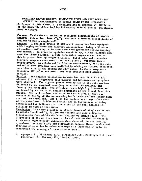
LOCALIZED PROTON DENSITY. RELAXATION TIMES AND SELF DIFFUSION<br />
COEFFICIENT MEASUREMENTS IN SINGLE CELLS BY NMR MICROSCOPY.<br />
J. AEuayo, S. Blackband, J. Schoenlger and M. Mattlngly*, Division<br />
of NMR Research, Johns Hopkins University Medical School, Baltlmore,<br />
Maryland 21205.<br />
l~urDose. To obtain and intrepret localised measurements of proton<br />
density, relaxation times (T1/T2, and self diffusion coefficients of<br />
regions wi<strong>th</strong>in a single cell.<br />
Me<strong>th</strong>ods. A modified Bruker AM 400 spectrometer has been equipped<br />
wi<strong>th</strong> imaging software and hardware accessories. Using a 50 mm set<br />
of gradient coils up to 20 G/cm have been generated during imaging<br />
experiments. In order to optimize sensitivity, a 5 mm solenoid were<br />
used for <strong>th</strong>ese studies. A spin echo pulse sequence was used to<br />
obtain proton density weighted images. Multi-echo and inversion<br />
recovery programs were used to obtain T 2 and T 1 weighted images<br />
respectively. To obtain self diffusion measurements, <strong>th</strong>e spin echo<br />
and multi-echo programs were modified by adding two pulsed gradients<br />
on ei<strong>th</strong>er side of <strong>th</strong>e refocusing 180 ° pulse. In <strong>th</strong>ese programs a<br />
selective 90 ° pulse was used. Ova were obtained from Xenopus<br />
laevius.<br />
Results. The highest resolution to date has been I0 X 13 X 250<br />
microns (I). A homogeneous cell nucleus and heterogenous cytoplasm<br />
were observed. The highest proton density was in <strong>th</strong>e cell nucleus<br />
followed by <strong>th</strong>e marginal zone (region around <strong>th</strong>e nucleus) and<br />
finally <strong>th</strong>e cytoplasm. The cytoplasm has a high lipid content as<br />
evidenced by a chemically shifted component of <strong>th</strong>e signal from <strong>th</strong>is<br />
region. The cell nuclues was noted to have a long T 1 <strong>th</strong>at was<br />
similar to <strong>th</strong>e T 1 of <strong>th</strong>e surrounding buffer solution and longer <strong>th</strong>an<br />
<strong>th</strong>at of <strong>th</strong>e cytoplasm. The T 2 of <strong>th</strong>e nucleus was longer <strong>th</strong>an <strong>th</strong>at<br />
of <strong>th</strong>e cytoplasm. Diffusion studies are in <strong>th</strong>e process of being<br />
Intrepreted but indicate <strong>th</strong>at <strong>th</strong>e water in <strong>th</strong>e cell nucleus is<br />
similiar to <strong>th</strong>at of free water.<br />
Conclusion. It is now possible to obtain images of single cells and<br />
to obtain localized TI, T2, proton density and self diffusion<br />
measurements from wi<strong>th</strong>in different regions of single cells. The<br />
properties of <strong>th</strong>e cell nucleus in <strong>th</strong>e cell system <strong>th</strong>at we chose to<br />
study were significantly different <strong>th</strong>an <strong>th</strong>ose of <strong>th</strong>e surrounding<br />
cytoplasm. Fur<strong>th</strong>er study and correlation between <strong>th</strong>ese results and<br />
previous observation by o<strong>th</strong>er techniques is important in order to<br />
understand <strong>th</strong>e meaning of <strong>th</strong>ese observations.<br />
I. Aguayo J.B., Blackband S.J., Schoeniger J.S., Mattingly M.O., and<br />
Hintermann M., Nature, 322, 190-192, 1986.


