

Acute Leukemias - Republican Scientific Medical Library
Acute Leukemias - Republican Scientific Medical Library
Acute Leukemias - Republican Scientific Medical Library

Create successful ePaper yourself
Turn your PDF publications into a flip-book with our unique Google optimized e-Paper software.
a 3.2 · Etiology 51<br />
part of the increase in median survival in the last years<br />
may be attributed to improved supportive care over past<br />
decades.<br />
3.1.7 Survivorship II<br />
At St. Jude Children’s Hospital, the incidence of and risk<br />
factors for the development of late sequelae of treatment<br />
in patients who survived for more than 10 years (median:<br />
15 years) after diagnosis of childhood AML have<br />
been evaluated. The most common late effects in adulthood<br />
consisted in growth abnormalities (51%). Depending<br />
on the treatment modality (chemotherapy only;<br />
combined chemo-, radiotherapy; or combined chemo-,<br />
radiotherapy with consecutive bone marrow or peripheral<br />
stem cell transplantation), endocrine abnormalities,<br />
cataracts, cardiac abnormalities, academic difficulties,<br />
and secondary malignancies resulted in 14–51%. Besides<br />
physical late effects, psychosocial complications were<br />
observed in long-term survivors [32].<br />
Patients that survived AML and treatment have also<br />
been monitored in a long-term follow-up at the University<br />
of Texas M.D. Anderson Cancer Center [33]. Some<br />
very relevant conclusions have been drawn in this report:<br />
Only 10% of all 1892 patients entered the potentially<br />
cured cohort, which was defined as the patient<br />
population in complete remission after a follow-up of<br />
3 years. Those patients in the potentially cured cohort<br />
were most likely to be able to return to work, suggesting<br />
that the major threat to patients with newly diagnosed<br />
AML is the disease and not the treatment.<br />
3.2 Etiology<br />
The development of AML has been associated with several<br />
risk factors. Remarkably though, as of yet defined<br />
risk factors account for only a small number of observed<br />
cases [34]. These include age, antecedent hematological<br />
disease, genetic disorders as well as exposures to<br />
viruses, radiation, chemical or other occupational hazards,<br />
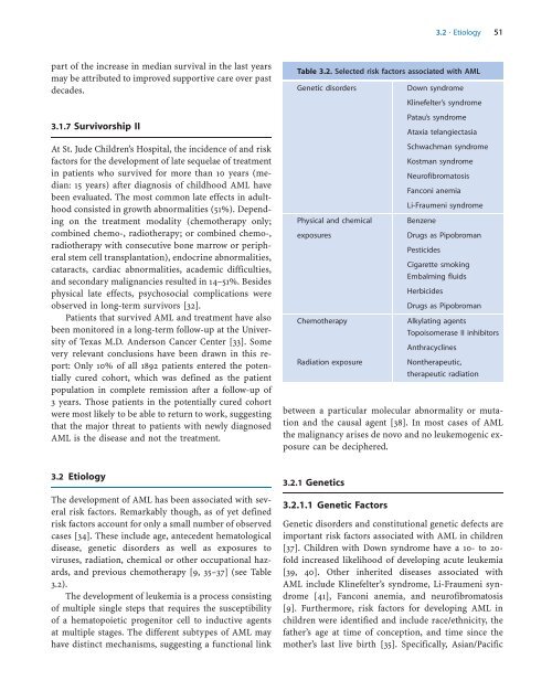
and previous chemotherapy [9, 35–37] (see Table<br />
3.2).<br />
The development of leukemia is a process consisting<br />
of multiple single steps that requires the susceptibility<br />
of a hematopoietic progenitor cell to inductive agents<br />
at multiple stages. The different subtypes of AML may<br />
have distinct mechanisms, suggesting a functional link<br />
Table 3.2. Selected risk factors associated with AML<br />
Genetic disorders Down syndrome<br />
Klinefelter’s syndrome<br />
Patau’s syndrome<br />
Ataxia telangiectasia<br />
Schwachman syndrome<br />
Kostman syndrome<br />
Neurofibromatosis<br />
Fanconi anemia<br />
Li-Fraumeni syndrome<br />
Physical and chemical Benzene<br />
exposures Drugs as Pipobroman<br />
Pesticides<br />
Cigarette smoking<br />
Embalming fluids<br />
Herbicides<br />
Drugs as Pipobroman<br />
Chemotherapy Alkylating agents<br />
Topoisomerase II inhibitors<br />
Anthracyclines<br />
Radiation exposure Nontherapeutic,<br />
therapeutic radiation<br />
between a particular molecular abnormality or mutation<br />
and the causal agent [38]. In most cases of AML<br />
the malignancy arises de novo and no leukemogenic exposure<br />
can be deciphered.<br />
3.2.1 Genetics<br />
3.2.1.1 Genetic Factors<br />
Genetic disorders and constitutional genetic defects are<br />
important risk factors associated with AML in children<br />
[37]. Children with Down syndrome have a 10- to 20fold<br />
increased likelihood of developing acute leukemia<br />
[39, 40]. Other inherited diseases associated with<br />
AML include Klinefelter’s syndrome, Li-Fraumeni syndrome<br />
[41], Fanconi anemia, and neurofibromatosis<br />
[9]. Furthermore, risk factors for developing AML in<br />
children were identified and include race/ethnicity, the<br />
father’s age at time of conception, and time since the<br />
mother’s last live birth [35]. Specifically, Asian/Pacific


