

Colposcopy and Treatment of Cervical Intraepithelial Neoplasia - RHO
Colposcopy and Treatment of Cervical Intraepithelial Neoplasia - RHO
Colposcopy and Treatment of Cervical Intraepithelial Neoplasia - RHO

You also want an ePaper? Increase the reach of your titles
YUMPU automatically turns print PDFs into web optimized ePapers that Google loves.
Chapter 12<br />
a b c<br />
d e f<br />
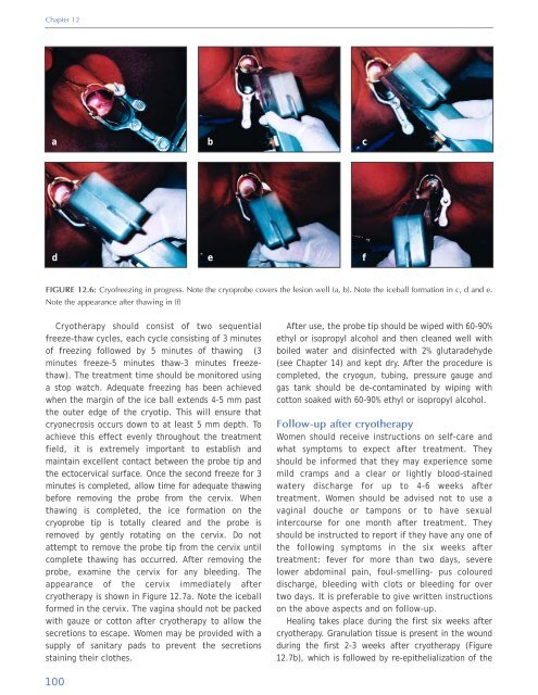
FIGURE 12.6: Cry<strong>of</strong>reezing in progress. Note the cryoprobe covers the lesion well (a, b). Note the iceball formation in c, d <strong>and</strong> e.<br />
Note the appearance after thawing in (f)<br />
Cryotherapy should consist <strong>of</strong> two sequential<br />
freeze-thaw cycles, each cycle consisting <strong>of</strong> 3 minutes<br />
<strong>of</strong> freezing followed by 5 minutes <strong>of</strong> thawing (3<br />
minutes freeze-5 minutes thaw-3 minutes freezethaw).<br />
The treatment time should be monitored using<br />
a stop watch. Adequate freezing has been achieved<br />
when the margin <strong>of</strong> the ice ball extends 4-5 mm past<br />
the outer edge <strong>of</strong> the cryotip. This will ensure that<br />
cryonecrosis occurs down to at least 5 mm depth. To<br />
achieve this effect evenly throughout the treatment<br />
field, it is extremely important to establish <strong>and</strong><br />
maintain excellent contact between the probe tip <strong>and</strong><br />
the ectocervical surface. Once the second freeze for 3<br />
minutes is completed, allow time for adequate thawing<br />
before removing the probe from the cervix. When<br />
thawing is completed, the ice formation on the<br />
cryoprobe tip is totally cleared <strong>and</strong> the probe is<br />
removed by gently rotating on the cervix. Do not<br />
attempt to remove the probe tip from the cervix until<br />
complete thawing has occurred. After removing the<br />
probe, examine the cervix for any bleeding. The<br />
appearance <strong>of</strong> the cervix immediately after<br />
cryotherapy is shown in Figure 12.7a. Note the iceball<br />
formed in the cervix. The vagina should not be packed<br />
with gauze or cotton after cryotherapy to allow the<br />
secretions to escape. Women may be provided with a<br />
supply <strong>of</strong> sanitary pads to prevent the secretions<br />
staining their clothes.<br />
After use, the probe tip should be wiped with 60-90%<br />
ethyl or isopropyl alcohol <strong>and</strong> then cleaned well with<br />
boiled water <strong>and</strong> disinfected with 2% glutaradehyde<br />
(see Chapter 14) <strong>and</strong> kept dry. After the procedure is<br />
completed, the cryogun, tubing, pressure gauge <strong>and</strong><br />
gas tank should be de-contaminated by wiping with<br />
cotton soaked with 60-90% ethyl or isopropyl alcohol.<br />
Follow-up after cryotherapy<br />
Women should receive instructions on self-care <strong>and</strong><br />
what symptoms to expect after treatment. They<br />
should be informed that they may experience some<br />
mild cramps <strong>and</strong> a clear or lightly blood-stained<br />
watery discharge for up to 4-6 weeks after<br />
treatment. Women should be advised not to use a<br />
vaginal douche or tampons or to have sexual<br />
intercourse for one month after treatment. They<br />
should be instructed to report if they have any one <strong>of</strong><br />
the following symptoms in the six weeks after<br />
treatment: fever for more than two days, severe<br />
lower abdominal pain, foul-smelling- pus coloured<br />
discharge, bleeding with clots or bleeding for over<br />
two days. It is preferable to give written instructions<br />
on the above aspects <strong>and</strong> on follow-up.<br />
Healing takes place during the first six weeks after<br />
cryotherapy. Granulation tissue is present in the wound<br />
during the first 2-3 weeks after cryotherapy (Figure<br />
12.7b), which is followed by re-epithelialization <strong>of</strong> the<br />
100


