

Colposcopy and Treatment of Cervical Intraepithelial Neoplasia - RHO
Colposcopy and Treatment of Cervical Intraepithelial Neoplasia - RHO
Colposcopy and Treatment of Cervical Intraepithelial Neoplasia - RHO

Create successful ePaper yourself
Turn your PDF publications into a flip-book with our unique Google optimized e-Paper software.
Chapter 4<br />
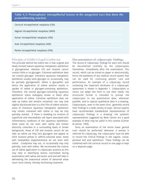
Table 4.2: Preneoplastic intraepithelial lesions in the anogenital tract that show the<br />
acetowhitening reaction<br />
<strong>Cervical</strong> intraepithelial neoplasia (CIN)<br />
Vaginal intraepithelial neoplasia (VAIN)<br />
Vulvar intraepithelial neoplasia (VIN)<br />
Anal intraepithelial neoplasia (AIN)<br />
Penile intraepithelial neoplasia (PIN)<br />
Principles <strong>of</strong> Schiller’s (Lugol’s) iodine test<br />
The principle behind the iodine test is that original <strong>and</strong><br />
newly formed mature squamous metaplastic epithelium<br />
is glycogenated, whereas CIN <strong>and</strong> invasive cancer<br />
contain little or no glycogen. Columnar epithelium does<br />
not contain glycogen. Immature squamous metaplastic<br />
epithelium usually lacks glycogen or, occasionally, may<br />
be partially glycogenated. Iodine is glycophilic <strong>and</strong><br />
hence the application <strong>of</strong> iodine solution results in<br />
uptake <strong>of</strong> iodine in glycogen-containing epithelium.<br />
Therefore, the normal glycogen-containing squamous<br />
epithelium stains mahogany brown or black after<br />
application <strong>of</strong> iodine. Columnar epithelium does not<br />
take up iodine <strong>and</strong> remains unstained, but may look<br />
slightly discoloured due to a thin film <strong>of</strong> iodine solution;<br />
areas <strong>of</strong> immature squamous metaplastic epithelium<br />
may remain unstained with iodine or may be only<br />
partially stained. If there is shedding (or erosion) <strong>of</strong><br />
superficial <strong>and</strong> intermediate cell layers associated with<br />
inflammatory conditions <strong>of</strong> the squamous epithelium,<br />
these areas do not stain with iodine <strong>and</strong> remain<br />
distinctly colourless in a surrounding black or brown<br />
background. Areas <strong>of</strong> CIN <strong>and</strong> invasive cancer do not<br />
take up iodine (as they lack glycogen) <strong>and</strong> appear as<br />
thick mustard yellow or saffron-coloured areas. Areas<br />
with leukoplakia (hyperkeratosis) do not stain with<br />
iodine. Condyloma may not, or occasionally may only<br />
partially, stain with iodine. We recommend the routine<br />
use <strong>of</strong> iodine application in colposcopic practice as this<br />
may help in identifying lesions overlooked during<br />
examination with saline <strong>and</strong> acetic acid <strong>and</strong> will help in<br />
delineating the anatomical extent <strong>of</strong> abnormal areas<br />
much more clearly, thereby facilitating treatment.<br />
Documentation <strong>of</strong> colposcopic findings<br />
The record <strong>of</strong> colposcopic findings for each visit should<br />
be documented carefully by the colposcopists<br />
themselves, immediately after the examination. This<br />
record, which can be stored on paper or electronically,<br />
forms the backbone <strong>of</strong> any medical record system that<br />
can be used for continuing patient care <strong>and</strong><br />
performance. An example <strong>of</strong> a colposcopy record<br />
containing the important attributes <strong>of</strong> a colposcopic<br />
assessment is shown in Appendix 1. Colposcopists or<br />
clinics can adapt this form to suit their needs; the<br />
structured format is intended to prompt the<br />
colposcopist to use quantitative data, whenever<br />
possible, <strong>and</strong> to capture qualitative data in a drawing.<br />
Colposcopists, even in the same clinic, generally record<br />
their findings in a wide variety <strong>of</strong> ways. Various experts<br />
have recommended st<strong>and</strong>ardized representations <strong>of</strong><br />
colposcopic findings in a drawing; the symbolic<br />
representations suggested by René Cartier are a good<br />
example <strong>of</strong> what may be useful in this context (Cartier<br />
& Cartier, 1993).<br />
Since an examination <strong>of</strong> the entire lower genital<br />
tract should be performed whenever a woman is<br />
referred for colposcopy, the colposcopist must be able<br />
to record the clinical findings in the vaginal, vulvar,<br />
perianal <strong>and</strong> anal epithelium. These findings can be<br />
combined with the cervical record on one page or kept<br />
on a separate page.<br />
36


