

Colposcopy and Treatment of Cervical Intraepithelial Neoplasia - RHO
Colposcopy and Treatment of Cervical Intraepithelial Neoplasia - RHO
Colposcopy and Treatment of Cervical Intraepithelial Neoplasia - RHO

You also want an ePaper? Increase the reach of your titles
YUMPU automatically turns print PDFs into web optimized ePapers that Google loves.
Colposcopic assessment <strong>of</strong> cervical intraepithelial neoplasia<br />
a<br />
c<br />
c<br />
b<br />
a<br />
FIGURE 7.19: A dense acetowhite lesion with varying colour<br />
intensity <strong>and</strong> coarse mosaics (a) in a CIN 2 lesion<br />
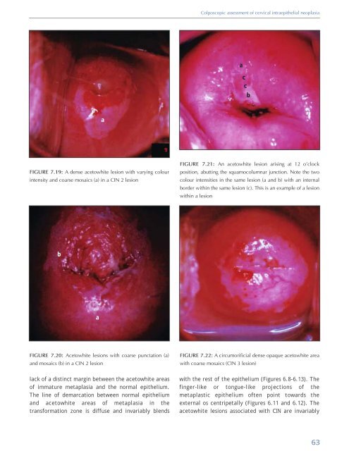
FIGURE 7.21: An acetowhite lesion arising at 12 o’clock<br />
position, abutting the squamocolumnar junction. Note the two<br />
colour intensities in the same lesion (a <strong>and</strong> b) with an internal<br />
border within the same lesion (c). This is an example <strong>of</strong> a lesion<br />
within a lesion<br />
b<br />
a<br />
FIGURE 7.20: Acetowhite lesions with coarse punctation (a)<br />
<strong>and</strong> mosaics (b) in a CIN 2 lesion<br />
FIGURE 7.22: A circumorificial dense opaque acetowhite area<br />
with coarse mosaics (CIN 3 lesion)<br />
lack <strong>of</strong> a distinct margin between the acetowhite areas<br />
<strong>of</strong> immature metaplasia <strong>and</strong> the normal epithelium.<br />
The line <strong>of</strong> demarcation between normal epithelium<br />
<strong>and</strong> acetowhite areas <strong>of</strong> metaplasia in the<br />
transformation zone is diffuse <strong>and</strong> invariably blends<br />
with the rest <strong>of</strong> the epithelium (Figures 6.8-6.13). The<br />
finger-like or tongue-like projections <strong>of</strong> the<br />
metaplastic epithelium <strong>of</strong>ten point towards the<br />
external os centripetally (Figures 6.11 <strong>and</strong> 6.12). The<br />
acetowhite lesions associated with CIN are invariably<br />
63


