

Colposcopy and Treatment of Cervical Intraepithelial Neoplasia - RHO
Colposcopy and Treatment of Cervical Intraepithelial Neoplasia - RHO
Colposcopy and Treatment of Cervical Intraepithelial Neoplasia - RHO

You also want an ePaper? Increase the reach of your titles
YUMPU automatically turns print PDFs into web optimized ePapers that Google loves.
An introduction to invasive cancer <strong>of</strong> the uterine cervix<br />
mushroom or proliferating, bulging cauliflower-like<br />
growth with polypoid or papillary excrescences.<br />
Endophytic cancers may extensively infiltrate the<br />
stroma, distorting the cervix, without much visible<br />
surface growth. These lesions may exp<strong>and</strong> into the<br />
endocervix leaving the squamous epithelium <strong>of</strong> the<br />
cervix intact until the lesions exceed 5-6 cm in a<br />
diameter. They result in a grossly enlarged, irregular<br />
barrel-shaped cervix with a rough, papillary or granular<br />
surface. Such cancers may remain silent for a long<br />
time. Partly exophytic <strong>and</strong> endophytic tumours are<br />
usually ulcerated with deep infiltration <strong>of</strong> the<br />
underlying stroma. In all types, bleeding on touch <strong>and</strong><br />
necrosis are predominant clinical features. Foulsmelling<br />
discharge is also common due to superadded<br />
anaerobic infection <strong>of</strong> the necrotic tissue.<br />
As the invasion continues further, it may directly<br />
involve the vagina, parametrium, pelvic sidewall,<br />
bladder <strong>and</strong> rectum. Compression <strong>of</strong> the ureter due to<br />
advanced local disease causes ureteral obstruction with<br />
resulting hydronephrosis (enlargement <strong>of</strong> kidneys) <strong>and</strong>,<br />
ultimately, renal failure. Regional lymph node<br />
metastasis occurs along with local invasion. Metastatic<br />
cancer in para-aortic nodes may extend through the<br />
node capsule <strong>and</strong> directly invade the vertebrae <strong>and</strong><br />
nerve roots. Direct invasion <strong>of</strong> the branches <strong>of</strong> the<br />
sciatic nerve roots causes back pain, <strong>and</strong> encroachment<br />
<strong>of</strong> the pelvic wall veins <strong>and</strong> lymphatics causes oedema<br />
<strong>of</strong> the lower limbs. Haematogenous spread to lumbar<br />
vertebrae <strong>and</strong> psoas muscle may occur without nodal<br />
disease. Distant metastases occur late in the disease,<br />
usually involving para-aortic nodes, lungs, liver, bone<br />
<strong>and</strong> other structures.<br />
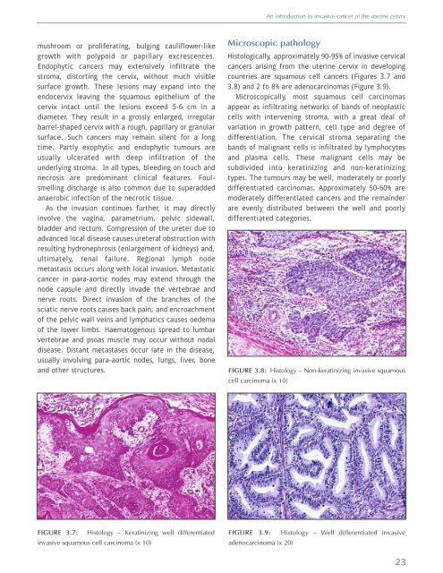
Microscopic pathology<br />
Histologically, approximately 90-95% <strong>of</strong> invasive cervical<br />
cancers arising from the uterine cervix in developing<br />
countries are squamous cell cancers (Figures 3.7 <strong>and</strong><br />
3.8) <strong>and</strong> 2 to 8% are adenocarcinomas (Figure 3.9).<br />
Microscopically, most squamous cell carcinomas<br />
appear as infiltrating networks <strong>of</strong> b<strong>and</strong>s <strong>of</strong> neoplastic<br />
cells with intervening stroma, with a great deal <strong>of</strong><br />
variation in growth pattern, cell type <strong>and</strong> degree <strong>of</strong><br />
differentiation. The cervical stroma separating the<br />
b<strong>and</strong>s <strong>of</strong> malignant cells is infiltrated by lymphocytes<br />
<strong>and</strong> plasma cells. These malignant cells may be<br />
subdivided into keratinizing <strong>and</strong> non-keratinizing<br />
types. The tumours may be well, moderately or poorly<br />
differentiated carcinomas. Approximately 50-60% are<br />
moderately differentiated cancers <strong>and</strong> the remainder<br />
are evenly distributed between the well <strong>and</strong> poorly<br />
differentiated categories.<br />
FIGURE 3.8: Histology – Non-keratinizing invasive squamous<br />
cell carcinoma (x 10)<br />
FIGURE 3.7: Histology – Keratinizing well differentiated<br />
invasive squamous cell carcinoma (x 10)<br />
FIGURE 3.9: Histology – Well differentiated invasive<br />
adenocarcinoma (x 20)<br />
23


