

Colposcopy and Treatment of Cervical Intraepithelial Neoplasia - RHO
Colposcopy and Treatment of Cervical Intraepithelial Neoplasia - RHO
Colposcopy and Treatment of Cervical Intraepithelial Neoplasia - RHO

You also want an ePaper? Increase the reach of your titles
YUMPU automatically turns print PDFs into web optimized ePapers that Google loves.
Chapter 5<br />
Obtain specimens for laboratory examination, if<br />
necessary<br />
Any necessary swab for screening or diagnostic work-up<br />
because <strong>of</strong> suspicious signs or symptoms should be done<br />
at this stage. For example, a swab for Neisseria<br />
gonorrhoeae culture can be obtained from the<br />
endocervical canal or pus in the vaginal fornix, <strong>and</strong> a<br />
Chlamydia trachomatis specimen can be obtained from<br />
the endocervical canal after excessive mucus has been<br />
removed. If an ulcerative lesion is found on the vagina<br />
or cervix or on the external anogenital area, the<br />
colposcopist should consider the possibility <strong>of</strong> one or<br />
more sexually transmitted infections as the cause <strong>and</strong><br />
the appropriate work up should be performed. If a<br />
sample is required to test for example for human<br />
papillomavirus (HPV), the cervical cells should be<br />
obtained before application <strong>of</strong> acetic acid.<br />
Following this, the cervix should be inspected at lowpower<br />
magnification (5x to 10x), looking for any<br />
obvious areas <strong>of</strong> abnormality (e.g., leukoplakia).<br />
Apply normal saline solution<br />
Normal saline is applied to the cervix with a sprayer or<br />
cotton balls <strong>and</strong> excess liquid is removed afterwards.<br />
This is not only the ideal way to conduct a preliminary<br />
inspection for surface abnormalities (e.g., leukoplakia,<br />
condylomata), but also the best way to examine the<br />
detail <strong>of</strong> cervical capillaries <strong>and</strong> surface blood vessels.<br />
The examination <strong>of</strong> the blood vessels is further aided<br />
by using the green (or blue) filter on the colposcope to<br />
enhance the contrast <strong>of</strong> the vessels, <strong>and</strong> by using<br />
higher levels <strong>of</strong> magnification (about 15x). Although<br />
some experienced colposcopists do not routinely<br />
perform an examination after saline has been applied<br />
(instead going directly to the application <strong>of</strong> acetic<br />
acid), it has been argued that an examination should be<br />
done in all cases, since the information obtained on the<br />
location <strong>of</strong> abnormal vessels can be noted <strong>and</strong><br />
integrated with the findings from later steps, which<br />
will determine the appropriate biopsy site(s), if any.<br />
The application <strong>of</strong> acetic acid, <strong>and</strong> even Lugol’s iodine<br />
solution, to the cervix can result in tissue swelling <strong>and</strong><br />
consequent opacity. This swelling <strong>and</strong> opacity tend to<br />
obscure some <strong>of</strong> the details <strong>of</strong> the vessels in the<br />
subepithelial tissue, so it is always is best to assess the<br />
capillaries <strong>and</strong> vessels with saline before the<br />
application <strong>of</strong> any other solution.<br />
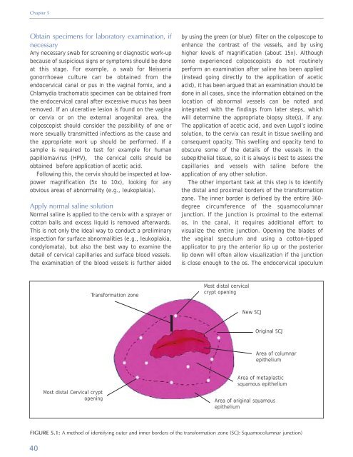
The other important task at this step is to identify<br />
the distal <strong>and</strong> proximal borders <strong>of</strong> the transformation<br />
zone. The inner border is defined by the entire 360-<br />
degree circumference <strong>of</strong> the squamocolumnar<br />
junction. If the junction is proximal to the external<br />
os, in the canal, it requires additional effort to<br />
visualize the entire junction. Opening the blades <strong>of</strong><br />
the vaginal speculum <strong>and</strong> using a cotton-tipped<br />
applicator to pry the anterior lip up or the posterior<br />
lip down will <strong>of</strong>ten allow visualization if the junction<br />
is close enough to the os. The endocervical speculum<br />
Transformation zone<br />
Most distal cervical<br />
crypt opening<br />
New SCJ<br />
Original SCJ<br />
Area <strong>of</strong> columnar<br />
epithelium<br />
Most distal <strong>Cervical</strong> crypt<br />
opening<br />
Area <strong>of</strong> metaplastic<br />
squamous epithelium<br />
Area <strong>of</strong> original squamous<br />
epithelium<br />
FIGURE 5.1: A method <strong>of</strong> identifying outer <strong>and</strong> inner borders <strong>of</strong> the transformation zone (SCJ: Squamocolumnar junction)<br />
40


