

Colposcopy and Treatment of Cervical Intraepithelial Neoplasia - RHO
Colposcopy and Treatment of Cervical Intraepithelial Neoplasia - RHO
Colposcopy and Treatment of Cervical Intraepithelial Neoplasia - RHO

You also want an ePaper? Increase the reach of your titles
YUMPU automatically turns print PDFs into web optimized ePapers that Google loves.
Colposcopic appearance <strong>of</strong> the normal cervix<br />
Afferent capillary<br />
FIGURE 6.4: Capillary network in columnar villi<br />
confined to the stromal core <strong>of</strong> each grape-like villus<br />
(Figure 6.4), which projects up to the epithelial surface.<br />
With the colposcope, the rounded tips <strong>of</strong> the individual<br />
villi are the main features seen <strong>and</strong> the top <strong>of</strong> the vessel<br />
network in each villus appears as a dot. Large, deep<br />
branching vessels may be seen in some cases.<br />
After application <strong>of</strong> 5% acetic acid solution<br />
Squamous epithelium<br />
After acetic acid has been allowed to take effect (1-2<br />
minutes), certain changes in the features seen with<br />
saline are usually apparent in the normal cervix <strong>of</strong> a<br />
young woman. The colour <strong>of</strong> the squamous epithelium<br />
tends to be somewhat dull in contrast to the usual pink<br />
hue, <strong>and</strong> the translucence is diminished so that it looks<br />
somewhat pale (Figure 6.1). In postmenopausal women<br />
the colour usually is paler than in a premenopausal<br />
woman. The l<strong>and</strong>marks <strong>and</strong> full extent <strong>of</strong> the<br />
Squamocolumnar<br />
juncton<br />
Columnar epithelium<br />
Efferent capillary<br />
Connective tissue<br />
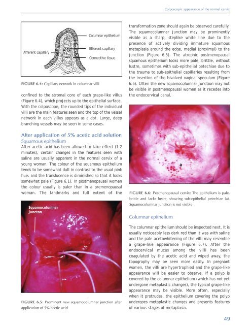
FIGURE 6.5: Prominent new squamocolumnar junction after<br />
application <strong>of</strong> 5% acetic acid<br />
transformation zone should again be observed carefully.<br />
The squamocolumnar junction may be prominently<br />
visible as a sharp, steplike white line due to the<br />
presence <strong>of</strong> actively dividing immature squamous<br />
metaplasia around the edge, medial (proximal) to the<br />
junction (Figure 6.5). The atrophic postmenopausal<br />
squamous epithelium looks more pale, brittle, without<br />
lustre, sometimes with sub-epithelial petechiae due to<br />
the trauma to sub-epithelial capillaries resulting from<br />
the insertion <strong>of</strong> the bivalved vaginal speculum (Figure<br />
6.6). Often the new squamocolumnar junction may not<br />
be visible in postmenopausal women as it recedes into<br />
the endocervical canal.<br />
FIGURE 6.6: Postmenopausal cervix: The epithelium is pale,<br />
brittle <strong>and</strong> lacks lustre, showing sub-epithelial petechiae (a).<br />
Squamocolumnar junction is not visible<br />
Columnar epithelium<br />
a<br />
The columnar epithelium should be inspected next. It is<br />
usually noticeably less dark red than it was with saline<br />
<strong>and</strong> the pale acetowhitening <strong>of</strong> the villi may resemble<br />
a grape-like appearance (Figure 6.7). After the<br />
endocervical mucus among the villi has been<br />
coagulated by the acetic acid <strong>and</strong> wiped away, the<br />
topography may be seen more easily. In pregnant<br />
women, the villi are hypertrophied <strong>and</strong> the grape-like<br />
appearance will be easier to observe. If a polyp is<br />
covered by the columnar epithelium (which has not yet<br />
undergone metaplastic changes), the typical grape-like<br />
appearance may be visible. More <strong>of</strong>ten, especially<br />
when it protrudes, the epithelium covering the polyp<br />
undergoes metaplastic changes <strong>and</strong> presents features<br />
<strong>of</strong> various stages <strong>of</strong> metaplasia.<br />
49


