

Colposcopy and Treatment of Cervical Intraepithelial Neoplasia - RHO
Colposcopy and Treatment of Cervical Intraepithelial Neoplasia - RHO
Colposcopy and Treatment of Cervical Intraepithelial Neoplasia - RHO

Create successful ePaper yourself
Turn your PDF publications into a flip-book with our unique Google optimized e-Paper software.
Chapter 1<br />
differentiation <strong>and</strong> maturation <strong>of</strong> these cells leads to<br />
the intermediate layers <strong>of</strong> polygonal cells with<br />
abundant cytoplasm <strong>and</strong> small round nuclei. These<br />
cells form a basket-weave pattern. With further<br />
maturation, the large <strong>and</strong> markedly flattened cells<br />
with small, dense, pyknotic nuclei <strong>and</strong> transparent<br />
cytoplasm <strong>of</strong> the superficial layers are formed.<br />
Overall, from the basal to the superficial layer, these<br />
cells undergo an increase in size <strong>and</strong> a reduction <strong>of</strong><br />
nuclear size.<br />
The intermediate <strong>and</strong> superficial layer cells<br />
contain abundant glycogen in their cytoplasm, which<br />
stains mahogany brown or black after application <strong>of</strong><br />
Lugol’s iodine <strong>and</strong> magenta with periodic acid-Schiff<br />
stain in histological sections. Glycogenation <strong>of</strong> the<br />
intermediate <strong>and</strong> superficial layers is a sign <strong>of</strong> normal<br />
maturation <strong>and</strong> development <strong>of</strong> the squamous<br />
epithelium. Abnormal or altered maturation is<br />
characterized by a lack <strong>of</strong> glycogen production.<br />
The maturation <strong>of</strong> the squamous epithelium <strong>of</strong> the<br />
cervix is dependent on estrogen, the female hormone.<br />
If estrogen is lacking, full maturation <strong>and</strong><br />
glycogenation does not take place. Hence, after<br />
menopause, the cells do not mature beyond the<br />
parabasal layer <strong>and</strong> do not accumulate as multiple<br />
layers <strong>of</strong> flat cells. Consequently, the epithelium<br />
becomes thin <strong>and</strong> atrophic. On visual examination, it<br />
appears pale, with subepithelial petechial<br />
haemorrhagic spots, as it is easily prone to trauma.<br />
Columnar epithelium<br />
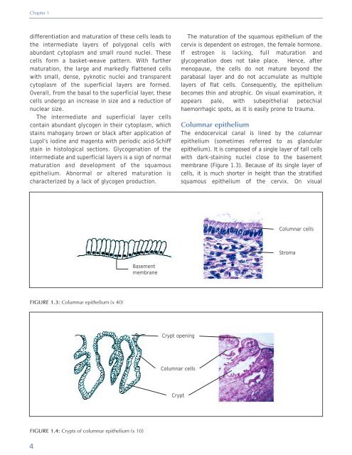
The endocervical canal is lined by the columnar<br />
epithelium (sometimes referred to as gl<strong>and</strong>ular<br />
epithelium). It is composed <strong>of</strong> a single layer <strong>of</strong> tall cells<br />
with dark-staining nuclei close to the basement<br />
membrane (Figure 1.3). Because <strong>of</strong> its single layer <strong>of</strong><br />
cells, it is much shorter in height than the stratified<br />
squamous epithelium <strong>of</strong> the cervix. On visual<br />
Columnar cells<br />
Stroma<br />
Basement<br />
membrane<br />
FIGURE 1.3: Columnar epithelium (x 40)<br />
Crypt opening<br />
Columnar cells<br />
Crypt<br />
FIGURE 1.4: Crypts <strong>of</strong> columnar epithelium (x 10)<br />
4


