

Colposcopy and Treatment of Cervical Intraepithelial Neoplasia - RHO
Colposcopy and Treatment of Cervical Intraepithelial Neoplasia - RHO
Colposcopy and Treatment of Cervical Intraepithelial Neoplasia - RHO

You also want an ePaper? Increase the reach of your titles
YUMPU automatically turns print PDFs into web optimized ePapers that Google loves.
Chapter 1<br />
Fundus<br />
Fallopian tube<br />
Body <strong>of</strong> uterus<br />
Supravaginal cervix<br />
Internal os<br />
Portio vaginalis<br />
Endocervical canal<br />
Endocervix<br />
Lateral fornix<br />
External os<br />
Ectocervix<br />
Vagina<br />
Cervix<br />
Bladder<br />
Anterior fornix<br />
Pubic bone<br />
Urethra<br />
Uterus<br />
Posterior fornix<br />
Rectum<br />
Sacrum<br />
Vagina<br />
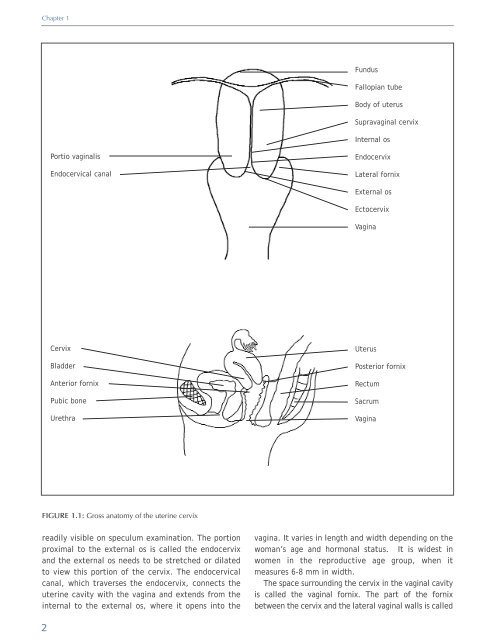
FIGURE 1.1: Gross anatomy <strong>of</strong> the uterine cervix<br />
readily visible on speculum examination. The portion<br />
proximal to the external os is called the endocervix<br />
<strong>and</strong> the external os needs to be stretched or dilated<br />
to view this portion <strong>of</strong> the cervix. The endocervical<br />
canal, which traverses the endocervix, connects the<br />
uterine cavity with the vagina <strong>and</strong> extends from the<br />
internal to the external os, where it opens into the<br />
vagina. It varies in length <strong>and</strong> width depending on the<br />
woman’s age <strong>and</strong> hormonal status. It is widest in<br />
women in the reproductive age group, when it<br />
measures 6-8 mm in width.<br />
The space surrounding the cervix in the vaginal cavity<br />
is called the vaginal fornix. The part <strong>of</strong> the fornix<br />
between the cervix <strong>and</strong> the lateral vaginal walls is called<br />
2


