

Colposcopy and Treatment of Cervical Intraepithelial Neoplasia - RHO
Colposcopy and Treatment of Cervical Intraepithelial Neoplasia - RHO
Colposcopy and Treatment of Cervical Intraepithelial Neoplasia - RHO

You also want an ePaper? Increase the reach of your titles
YUMPU automatically turns print PDFs into web optimized ePapers that Google loves.
<strong>Treatment</strong> <strong>of</strong> cervical intraepithelial neoplasia by loop electrosurgical excision procedure (LEEP)<br />
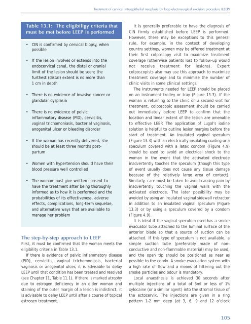
Table 13.1: The eligibiligy criteria that<br />
must be met before LEEP is performed<br />
• CIN is confirmed by cervical biopsy, when<br />
possible<br />
• If the lesion involves or extends into the<br />
endocervical canal, the distal or cranial<br />
limit <strong>of</strong> the lesion should be seen; the<br />
furthest (distal) extent is no more than<br />
1 cm in depth<br />
• There is no evidence <strong>of</strong> invasive cancer or<br />
gl<strong>and</strong>ular dysplasia<br />
• There is no evidence <strong>of</strong> pelvic<br />
inflammatory disease (PID), cervicitis,<br />
vaginal trichomoniasis, bacterial vaginosis,<br />
anogenital ulcer or bleeding disorder<br />
• If the woman has recently delivered, she<br />
should be at least three months postpartum<br />
• Women with hypertension should have their<br />
blood pressure well controlled<br />
• The woman must give written consent to<br />
have the treatment after being thoroughly<br />
informed as to how it is performed <strong>and</strong> the<br />
probabilities <strong>of</strong> its effectiveness, adverse<br />
effects, complications, long-term sequelae,<br />
<strong>and</strong> alternative ways that are available to<br />
manage her problem<br />
The step-by-step approach to LEEP<br />
First, it must be confirmed that the woman meets the<br />
eligibility criteria in Table 13.1.<br />
If there is evidence <strong>of</strong> pelvic inflammatory disease<br />
(PID), cervicitis, vaginal trichomoniasis, bacterial<br />
vaginosis or anogenital ulcer, it is advisable to delay<br />
LEEP until that condition has been treated <strong>and</strong> resolved<br />
(see Chapter 11, Table 11.1). If there is marked atrophy<br />
due to estrogen deficiency in an older woman <strong>and</strong><br />
staining <strong>of</strong> the outer margin <strong>of</strong> a lesion is indistinct, it<br />
is advisable to delay LEEP until after a course <strong>of</strong> topical<br />
estrogen treatment.<br />
It is generally preferable to have the diagnosis <strong>of</strong><br />
CIN firmly established before LEEP is performed.<br />
However, there may be exceptions to this general<br />
rule, for example, in the context <strong>of</strong> developing<br />
country settings, women may be <strong>of</strong>fered treatment at<br />
their first colposcopy visit to maximize treatment<br />
coverage (otherwise patients lost to follow-up would<br />
not receive treatment for lesions). Expert<br />
colposcopists also may use this approach to maximize<br />
treatment coverage <strong>and</strong> to minimise the number <strong>of</strong><br />
clinic visits in some clinical settings.<br />
The instruments needed for LEEP should be placed<br />
on an instrument trolley or tray (Figure 13.3). If the<br />
woman is returning to the clinic on a second visit for<br />
treatment, colposcopic assessment should be carried<br />
out immediately before LEEP to confirm that the<br />
location <strong>and</strong> linear extent <strong>of</strong> the lesion are amenable<br />
to effective LEEP. The application <strong>of</strong> Lugol’s iodine<br />
solution is helpful to outline lesion margins before the<br />
start <strong>of</strong> treatment. An insulated vaginal speculum<br />
(Figure 13.3) with an electrically insulating coating or a<br />
speculum covered with a latex condom (Figure 4.9)<br />
should be used to avoid an electrical shock to the<br />
woman in the event that the activated electrode<br />
inadvertently touches the speculum (though this type<br />
<strong>of</strong> event usually does not cause any tissue damage<br />
because <strong>of</strong> the relatively large area <strong>of</strong> contact).<br />
Similarly, care must be taken to avoid causing pain by<br />
inadvertently touching the vaginal walls with the<br />
activated electrode. The later possibility may be<br />
avoided by using an insulated vaginal sidewall retractor<br />
in addition to an insulated vaginal speculum (Figure<br />
13.3) or by using a speculum covered by a condom<br />
(Figure 4.9).<br />
It is ideal if the vaginal speculum used has a smoke<br />
evacuator tube attached to the luminal surface <strong>of</strong> the<br />
anterior blade so that a source <strong>of</strong> suction can be<br />
attached. If this type <strong>of</strong> speculum is not available, a<br />
simple suction tube (preferably made <strong>of</strong> nonconductive<br />
<strong>and</strong> non-flammable material) may be used,<br />
<strong>and</strong> the open tip should be positioned as near as<br />
possible to the cervix. A smoke evacuation system with<br />
a high rate <strong>of</strong> flow <strong>and</strong> a means <strong>of</strong> filtering out the<br />
smoke particles <strong>and</strong> odour is m<strong>and</strong>atory.<br />
Local anaesthesia is achieved 30 seconds after<br />
multiple injections <strong>of</strong> a total <strong>of</strong> 5ml or less <strong>of</strong> 1%<br />
xylocaine (or a similar agent) into the stromal tissue <strong>of</strong><br />
the ectocervix. The injections are given in a ring<br />
pattern 1-2 mm deep (at 3, 6, 9 <strong>and</strong> 12 o’clock<br />
105


