

Colposcopy and Treatment of Cervical Intraepithelial Neoplasia - RHO
Colposcopy and Treatment of Cervical Intraepithelial Neoplasia - RHO
Colposcopy and Treatment of Cervical Intraepithelial Neoplasia - RHO

Create successful ePaper yourself
Turn your PDF publications into a flip-book with our unique Google optimized e-Paper software.
Chapter 9<br />
involving the columnar epithelium <strong>of</strong> the cervix. It<br />
results in congestion <strong>of</strong> underlying connective tissue,<br />
desquamation <strong>of</strong> cells <strong>and</strong> ulceration with<br />
mucopurulent discharge. If the inflammation persists,<br />
the villous structures become flattened, <strong>and</strong> the<br />
grape-like appearance is lost <strong>and</strong> the mucosa may<br />
secrete less mucus.<br />
In both the above conditions, after repeated<br />
inflammation <strong>and</strong> tissue necrosis, the lesions are<br />
repaired <strong>and</strong> necrotic tissue is eliminated. The newly<br />
formed epithelium has numerous vessels, <strong>and</strong><br />
connective tissue proliferation results in fibrosis <strong>of</strong><br />
varying extent.<br />
Colposcopic appearances<br />
Before the application <strong>of</strong> acetic acid<br />
Examination, before application <strong>of</strong> acetic acid,<br />
reveals moderate to excessive cervical <strong>and</strong> vaginal<br />
secretions, which may sometimes indicate the nature<br />
<strong>of</strong> underlying infection. In T. vaginalis infection<br />
(trichomoniasis), which is very common in tropical<br />
areas, there is copious, bubbly, frothy, malodorous,<br />
greenish-yellow, mucopurulent discharge. Bacterial<br />
infections are associated with thin, liquid,<br />
seropurulent discharge. The secretion may be foulsmelling<br />
in the case <strong>of</strong> anaerobic bacterial<br />
overgrowth, bacterial vaginosis, <strong>and</strong> Trichomonas<br />
infection. In the case <strong>of</strong> c<strong>and</strong>idiasis (moniliasis) <strong>and</strong><br />
other yeast infections, the secretion is thick <strong>and</strong> curdy<br />
(cheesy) white with intense itching resulting in a<br />
reddened vulva. Foul-smelling, dark-coloured<br />
mucopurulent discharges are associated with<br />
inflammatory states due to foreign bodies (e.g., a<br />
retained tampon). Gonorrhoea results in purulent<br />
vaginal discharge <strong>and</strong> cervical tenderness. Small<br />
vesicles filled with serous fluid may be observed in the<br />
cervix <strong>and</strong> vagina in the vesicular phase <strong>of</strong> herpes<br />
simplex viral infection. Herpetic infections are<br />
associated with episodes <strong>of</strong> painful vulvar, vaginal <strong>and</strong><br />
cervical ulceration lasting for two weeks. Excoriation<br />
marks are evident with trichomoniasis, moniliasis <strong>and</strong><br />
mixed bacterial infections.<br />
A large coalesced ulcer due to herpes, or other<br />
inflammatory conditions, may mimic the appearance<br />
<strong>of</strong> invasive cancer. Chronic inflammation may cause<br />
recurrent ulceration <strong>and</strong> healing <strong>of</strong> the cervix,<br />
resulting in distortion <strong>of</strong> the cervix due to healing by<br />
fibrosis. There may be associated necrotic areas as<br />
well. A biopsy should be directed if in doubt. Rare <strong>and</strong><br />
uncommon cervical infections, due to tuberculosis,<br />
schistosomiasis <strong>and</strong> amoebiasis, cause extensive<br />
ulceration <strong>and</strong> necrosis <strong>of</strong> the cervix with symptoms<br />
<strong>and</strong> signs mimicking invasive cancer; a biopsy will<br />
confirm the diagnosis.<br />
If the infectious process is accompanied by marked<br />
ulceration (with or without necrosis), the ulcerated<br />
area may be covered with purulent exudate, with<br />
marked differences in the surface level <strong>of</strong> the cervix.<br />
There may be exudation <strong>of</strong> serous droplets.<br />
Longst<strong>and</strong>ing bacterial, fungal or protozoal<br />
infection <strong>and</strong> inflammation may lead to fibrosis, which<br />
appears white or pink, depending on the degree <strong>of</strong><br />
fibrosis. The epithelium covering the connective tissue<br />
is fragile, leading to ulceration <strong>and</strong> bleeding.<br />
Appearances following acetic acid <strong>and</strong> iodine<br />
application are variable, depending on the integrity <strong>of</strong><br />
the surface epithelium.<br />
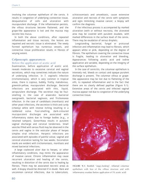
In the case <strong>of</strong> cervicitis, the columnar epithelium is<br />
intensely red, bleeds on contact <strong>and</strong> opaque purulent<br />
discharge is present. The columnar villous or grapelike<br />
appearance may be lost due to flattening <strong>of</strong> the<br />
villi, to repeated inflammation <strong>and</strong> to the fact that<br />
there are no clearly defined papillae (Figure 9.1).<br />
Extensive areas <strong>of</strong> the cervix <strong>and</strong> infected vaginal<br />
mucosa appear red due to congestion <strong>of</strong> the underlying<br />
connective tissue.<br />
FIGURE 9.1: Reddish “angry-looking”, inflamed columnar<br />
epithelium with loss <strong>of</strong> the villous structure <strong>and</strong> with<br />
inflammatory exudate (before application <strong>of</strong> 5% acetic acid)<br />
80


