

Colposcopy and Treatment of Cervical Intraepithelial Neoplasia - RHO
Colposcopy and Treatment of Cervical Intraepithelial Neoplasia - RHO
Colposcopy and Treatment of Cervical Intraepithelial Neoplasia - RHO

Create successful ePaper yourself
Turn your PDF publications into a flip-book with our unique Google optimized e-Paper software.
Chapter 4<br />
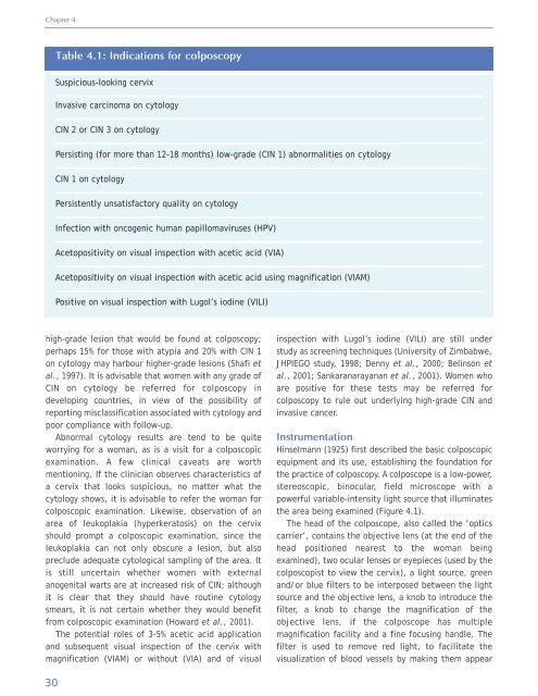
Table 4.1: Indications for colposcopy<br />
Suspicious-looking cervix<br />
Invasive carcinoma on cytology<br />
CIN 2 or CIN 3 on cytology<br />
Persisting (for more than 12-18 months) low-grade (CIN 1) abnormalities on cytology<br />
CIN 1 on cytology<br />
Persistently unsatisfactory quality on cytology<br />
Infection with oncogenic human papillomaviruses (HPV)<br />
Acetopositivity on visual inspection with acetic acid (VIA)<br />
Acetopositivity on visual inspection with acetic acid using magnification (VIAM)<br />
Positive on visual inspection with Lugol’s iodine (VILI)<br />
high-grade lesion that would be found at colposcopy;<br />
perhaps 15% for those with atypia <strong>and</strong> 20% with CIN 1<br />
on cytology may harbour higher-grade lesions (Shafi et<br />
al., 1997). It is advisable that women with any grade <strong>of</strong><br />
CIN on cytology be referred for colposcopy in<br />
developing countries, in view <strong>of</strong> the possibility <strong>of</strong><br />
reporting misclassification associated with cytology <strong>and</strong><br />
poor compliance with follow-up.<br />
Abnormal cytology results are tend to be quite<br />
worrying for a woman, as is a visit for a colposcopic<br />
examination. A few clinical caveats are worth<br />
mentioning. If the clinician observes characteristics <strong>of</strong><br />
a cervix that looks suspicious, no matter what the<br />
cytology shows, it is advisable to refer the woman for<br />
colposcopic examination. Likewise, observation <strong>of</strong> an<br />
area <strong>of</strong> leukoplakia (hyperkeratosis) on the cervix<br />
should prompt a colposcopic examination, since the<br />
leukoplakia can not only obscure a lesion, but also<br />
preclude adequate cytological sampling <strong>of</strong> the area. It<br />
is still uncertain whether women with external<br />
anogenital warts are at increased risk <strong>of</strong> CIN; although<br />
it is clear that they should have routine cytology<br />
smears, it is not certain whether they would benefit<br />
from colposcopic examination (Howard et al., 2001).<br />
The potential roles <strong>of</strong> 3-5% acetic acid application<br />
<strong>and</strong> subsequent visual inspection <strong>of</strong> the cervix with<br />
magnification (VIAM) or without (VIA) <strong>and</strong> <strong>of</strong> visual<br />
inspection with Lugol’s iodine (VILI) are still under<br />
study as screening techniques (University <strong>of</strong> Zimbabwe,<br />
JHPIEGO study, 1998; Denny et al., 2000; Belinson et<br />
al., 2001; Sankaranarayanan et al., 2001). Women who<br />
are positive for these tests may be referred for<br />
colposcopy to rule out underlying high-grade CIN <strong>and</strong><br />
invasive cancer.<br />
Instrumentation<br />
Hinselmann (1925) first described the basic colposcopic<br />
equipment <strong>and</strong> its use, establishing the foundation for<br />
the practice <strong>of</strong> colposcopy. A colposcope is a low-power,<br />
stereoscopic, binocular, field microscope with a<br />
powerful variable-intensity light source that illuminates<br />
the area being examined (Figure 4.1).<br />
The head <strong>of</strong> the colposcope, also called the ‘optics<br />
carrier’, contains the objective lens (at the end <strong>of</strong> the<br />
head positioned nearest to the woman being<br />
examined), two ocular lenses or eyepieces (used by the<br />
colposcopist to view the cervix), a light source, green<br />
<strong>and</strong>/or blue filters to be interposed between the light<br />
source <strong>and</strong> the objective lens, a knob to introduce the<br />
filter, a knob to change the magnification <strong>of</strong> the<br />
objective lens, if the colposcope has multiple<br />
magnification facility <strong>and</strong> a fine focusing h<strong>and</strong>le. The<br />
filter is used to remove red light, to facilitate the<br />
visualization <strong>of</strong> blood vessels by making them appear<br />
30


