

Colposcopy and Treatment of Cervical Intraepithelial Neoplasia - RHO
Colposcopy and Treatment of Cervical Intraepithelial Neoplasia - RHO
Colposcopy and Treatment of Cervical Intraepithelial Neoplasia - RHO

You also want an ePaper? Increase the reach of your titles
YUMPU automatically turns print PDFs into web optimized ePapers that Google loves.
Chapter 13<br />
Once the specimen has been removed <strong>and</strong> placed in<br />
formalin, the setting on the electrosurgical generator is<br />
changed to fulguration <strong>and</strong> the appropriate power is<br />
selected. The surface <strong>of</strong> the excisional crater is<br />
fulgurated using 3 or 5 mm ball electrode, in the<br />
coagulation mode. The edges <strong>of</strong> the crater should also<br />
be fulgurated to preserve the squamocolumnar junction<br />
in the visible ectocervix. If active bleeding occurs <strong>and</strong><br />
is difficult to control using the ball electrode, a<br />
macroneedle style electrode can be effectively used to<br />
apply the fulguration current in a much more<br />
concentrated (higher current density) <strong>and</strong> localized<br />
fashion to a bleeding site. If satisfactory haemostasis<br />
has been obtained, the surface <strong>of</strong> the crater is then<br />
coated with Monsel’s paste <strong>and</strong> the speculum is<br />
removed. It is a general observation that an extremely<br />
nervous patient tends to bleed more than a relaxed one<br />
- another good reason to communicate with the patient<br />
throughout the procedure <strong>and</strong> to try to calm her fears.<br />
If bleeding is difficult to stop despite use <strong>of</strong> the<br />
methods outlined above, the base <strong>of</strong> the excisional<br />
crater should be liberally coated with Monsel’s paste<br />
<strong>and</strong> the vagina packed with gauze. The woman should<br />
be asked to wait for several hours before removing the<br />
pack. This complication appears to occur more<br />
frequently in women with cervicitis.<br />
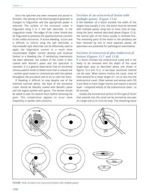
Excision <strong>of</strong> an ectocervical lesion with<br />
multiple passes (Figure 13.6)<br />
If the diameter <strong>of</strong> a lesion exceeds the width <strong>of</strong> the<br />
largest loop (usually 2 cm), the lesion must be removed<br />
with multiple passes using one or more sizes <strong>of</strong> loop.<br />
Using the basic method described above (Figure 13.3),<br />
the central part <strong>of</strong> the lesion usually is removed first.<br />
The remaining parts <strong>of</strong> the lesion in the periphery are<br />
then removed by one or more separate passes. All<br />
specimens are preserved for pathological examination.<br />
Excision <strong>of</strong> ectocervical plus endocervical<br />
lesions (Figures 13.7 <strong>and</strong> 13.8)<br />
If a lesion involves the endocervical canal <strong>and</strong> is not<br />
likely to be removed with the depth <strong>of</strong> the usual<br />
single-layer pass as described above <strong>and</strong> shown in<br />
figures 13.4 <strong>and</strong> 13.5, a two-layer excisional method<br />
can be used. When lesions involve the canal, most <strong>of</strong><br />
them extend for a linear length <strong>of</strong> 1 cm or less into the<br />
endocervical canal. Older women <strong>and</strong> women with CIN<br />
3 are likely to have longer lesions <strong>and</strong> require a second<br />
layer - composed wholly <strong>of</strong> the endocervical canal - to<br />
be excised.<br />
Usually the ectocervical portion <strong>of</strong> this type <strong>of</strong> lesion<br />
that extends into the canal can be excised by one pass<br />
<strong>of</strong> a large oval (2.0 x 0.8 cm) loop. The remaining tissue<br />
FIGURE 13.6: Excision <strong>of</strong> an ectocervical lesion with multiple passes<br />
108


