

Colposcopy and Treatment of Cervical Intraepithelial Neoplasia - RHO
Colposcopy and Treatment of Cervical Intraepithelial Neoplasia - RHO
Colposcopy and Treatment of Cervical Intraepithelial Neoplasia - RHO

You also want an ePaper? Increase the reach of your titles
YUMPU automatically turns print PDFs into web optimized ePapers that Google loves.
Chapter 8<br />
diagnosis <strong>of</strong> AIS <strong>and</strong> adenocarcinoma require a high<br />
degree <strong>of</strong> training <strong>and</strong> skill.<br />
It has been suggested that most gl<strong>and</strong>ular lesions<br />
originate within the transformation zone <strong>and</strong><br />
colposcopic recognition <strong>of</strong> the stark acetowhiteness <strong>of</strong><br />
either the individual or fused villi in discrete patches<br />
(in contrast to the surrounding pinkish white columnar<br />
villi) may lead to a colposcopic suspicion <strong>of</strong> gl<strong>and</strong>ular<br />
lesions. While CIN lesions are almost always<br />
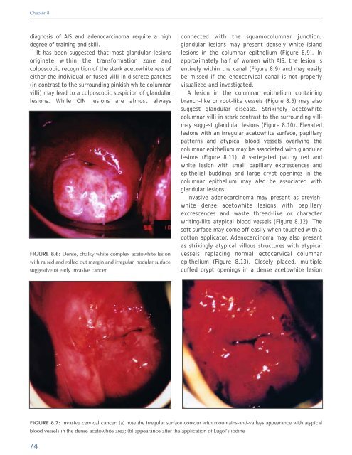
FIGURE 8.6: Dense, chalky white complex acetowhite lesion<br />
with raised <strong>and</strong> rolled out margin <strong>and</strong> irregular, nodular surface<br />
suggestive <strong>of</strong> early invasive cancer<br />
connected with the squamocolumnar junction,<br />
gl<strong>and</strong>ular lesions may present densely white isl<strong>and</strong><br />
lesions in the columnar epithelium (Figure 8.9). In<br />
approximately half <strong>of</strong> women with AIS, the lesion is<br />
entirely within the canal (Figure 8.9) <strong>and</strong> may easily<br />
be missed if the endocervical canal is not properly<br />
visualized <strong>and</strong> investigated.<br />
A lesion in the columnar epithelium containing<br />
branch-like or root-like vessels (Figure 8.5) may also<br />
suggest gl<strong>and</strong>ular disease. Strikingly acetowhite<br />
columnar villi in stark contrast to the surrounding villi<br />
may suggest gl<strong>and</strong>ular lesions (Figure 8.10). Elevated<br />
lesions with an irregular acetowhite surface, papillary<br />
patterns <strong>and</strong> atypical blood vessels overlying the<br />
columnar epithelium may be associated with gl<strong>and</strong>ular<br />
lesions (Figure 8.11). A variegated patchy red <strong>and</strong><br />
white lesion with small papillary excrescences <strong>and</strong><br />
epithelial buddings <strong>and</strong> large crypt openings in the<br />
columnar epithelium may also be associated with<br />
gl<strong>and</strong>ular lesions.<br />
Invasive adenocarcinoma may present as greyishwhite<br />
dense acetowhite lesions with papillary<br />
excrescences <strong>and</strong> waste thread-like or character<br />
writing-like atypical blood vessels (Figure 8.12). The<br />
s<strong>of</strong>t surface may come <strong>of</strong>f easily when touched with a<br />
cotton applicator. Adenocarcinoma may also present<br />
as strikingly atypical villous structures with atypical<br />
vessels replacing normal ectocervical columnar<br />
epithelium (Figure 8.13). Closely placed, multiple<br />
cuffed crypt openings in a dense acetowhite lesion<br />
FIGURE 8.7: Invasive cervical cancer: (a) note the irregular surface contour with mountains-<strong>and</strong>-valleys appearance with atypical<br />
blood vessels in the dense acetowhite area; (b) appearance after the application <strong>of</strong> Lugol’s iodine<br />
74


