

Colposcopy and Treatment of Cervical Intraepithelial Neoplasia - RHO
Colposcopy and Treatment of Cervical Intraepithelial Neoplasia - RHO
Colposcopy and Treatment of Cervical Intraepithelial Neoplasia - RHO

You also want an ePaper? Increase the reach of your titles
YUMPU automatically turns print PDFs into web optimized ePapers that Google loves.
Colposcopic assessment <strong>of</strong> cervical intraepithelial neoplasia<br />
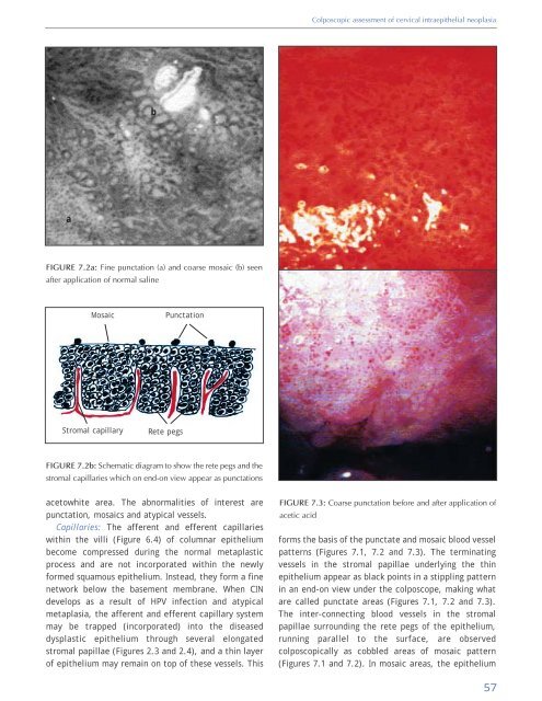
b<br />
a<br />
FIGURE 7.2a: Fine punctation (a) <strong>and</strong> coarse mosaic (b) seen<br />
after application <strong>of</strong> normal saline<br />
Mosaic<br />
Punctation<br />
Stromal capillary<br />
Rete pegs<br />
FIGURE 7.2b: Schematic diagram to show the rete pegs <strong>and</strong> the<br />
stromal capillaries which on end-on view appear as punctations<br />
acetowhite area. The abnormalities <strong>of</strong> interest are<br />
punctation, mosaics <strong>and</strong> atypical vessels.<br />
Capillaries: The afferent <strong>and</strong> efferent capillaries<br />
within the villi (Figure 6.4) <strong>of</strong> columnar epithelium<br />
become compressed during the normal metaplastic<br />
process <strong>and</strong> are not incorporated within the newly<br />
formed squamous epithelium. Instead, they form a fine<br />
network below the basement membrane. When CIN<br />
develops as a result <strong>of</strong> HPV infection <strong>and</strong> atypical<br />
metaplasia, the afferent <strong>and</strong> efferent capillary system<br />
may be trapped (incorporated) into the diseased<br />
dysplastic epithelium through several elongated<br />
stromal papillae (Figures 2.3 <strong>and</strong> 2.4), <strong>and</strong> a thin layer<br />
<strong>of</strong> epithelium may remain on top <strong>of</strong> these vessels. This<br />
FIGURE 7.3: Coarse punctation before <strong>and</strong> after application <strong>of</strong><br />
acetic acid<br />
forms the basis <strong>of</strong> the punctate <strong>and</strong> mosaic blood vessel<br />
patterns (Figures 7.1, 7.2 <strong>and</strong> 7.3). The terminating<br />
vessels in the stromal papillae underlying the thin<br />
epithelium appear as black points in a stippling pattern<br />
in an end-on view under the colposcope, making what<br />
are called punctate areas (Figures 7.1, 7.2 <strong>and</strong> 7.3).<br />
The inter-connecting blood vessels in the stromal<br />
papillae surrounding the rete pegs <strong>of</strong> the epithelium,<br />
running parallel to the surface, are observed<br />
colposcopically as cobbled areas <strong>of</strong> mosaic pattern<br />
(Figures 7.1 <strong>and</strong> 7.2). In mosaic areas, the epithelium<br />
57


