

Colposcopy and Treatment of Cervical Intraepithelial Neoplasia - RHO
Colposcopy and Treatment of Cervical Intraepithelial Neoplasia - RHO
Colposcopy and Treatment of Cervical Intraepithelial Neoplasia - RHO

Create successful ePaper yourself
Turn your PDF publications into a flip-book with our unique Google optimized e-Paper software.
Chapter 7<br />
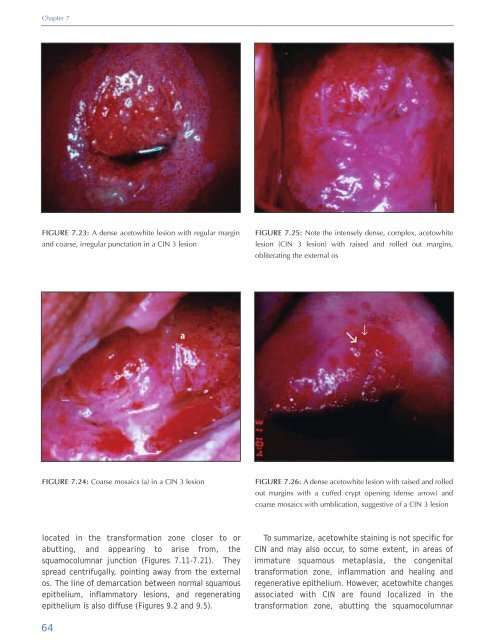
FIGURE 7.23: A dense acetowhite lesion with regular margin<br />
<strong>and</strong> coarse, irregular punctation in a CIN 3 lesion<br />
FIGURE 7.25: Note the intensely dense, complex, acetowhite<br />
lesion (CIN 3 lesion) with raised <strong>and</strong> rolled out margins,<br />
obliterating the external os<br />
a<br />
↑<br />
↑<br />
FIGURE 7.24: Coarse mosaics (a) in a CIN 3 lesion<br />
FIGURE 7.26: A dense acetowhite lesion with raised <strong>and</strong> rolled<br />
out margins with a cuffed crypt opening (dense arrow) <strong>and</strong><br />
coarse mosaics with umblication, suggestive <strong>of</strong> a CIN 3 lesion<br />
located in the transformation zone closer to or<br />
abutting, <strong>and</strong> appearing to arise from, the<br />
squamocolumnar junction (Figures 7.11-7.21). They<br />
spread centrifugally, pointing away from the external<br />
os. The line <strong>of</strong> demarcation between normal squamous<br />
epithelium, inflammatory lesions, <strong>and</strong> regenerating<br />
epithelium is also diffuse (Figures 9.2 <strong>and</strong> 9.5).<br />
To summarize, acetowhite staining is not specific for<br />
CIN <strong>and</strong> may also occur, to some extent, in areas <strong>of</strong><br />
immature squamous metaplasia, the congenital<br />
transformation zone, inflammation <strong>and</strong> healing <strong>and</strong><br />
regenerative epithelium. However, acetowhite changes<br />
associated with CIN are found localized in the<br />
transformation zone, abutting the squamocolumnar<br />
64


