

Colposcopy and Treatment of Cervical Intraepithelial Neoplasia - RHO
Colposcopy and Treatment of Cervical Intraepithelial Neoplasia - RHO
Colposcopy and Treatment of Cervical Intraepithelial Neoplasia - RHO

Create successful ePaper yourself
Turn your PDF publications into a flip-book with our unique Google optimized e-Paper software.
Colposcopic appearance <strong>of</strong> the normal cervix<br />
a<br />
↑<br />
FIGURE 6.10: The glassy, pinkishwhite immature squamous<br />
metaplastic epithelium (a) with isl<strong>and</strong>s <strong>of</strong> columnar epithelium<br />
(narrow arrow) <strong>and</strong> crypt opening (bold arrow) (after 5% acetic<br />
acid application)<br />
↑<br />
↑<br />
a<br />
c<br />
b<br />
the metaplastic process progresses. Gradually, the<br />
tongue-like metaplastic areas fuse together to form a<br />
continuously advancing glassy, shining, pinkish-white or<br />
mildly pale membrane-like area (Figure 6.13).<br />
Finally, the immature metaplastic epithelium<br />
becomes a fully developed mature metaplastic<br />
squamous epithelium resembling the original native<br />
squamous epithelium, except for the presence <strong>of</strong> some<br />
crypt openings (Figure 6.1) <strong>and</strong> nabothian retention<br />
follicles in the metaplastic epithelium (Figures 1.11,<br />
a<br />
b<br />
a<br />
FIGURE 6.11: The prominent white line corresponds to the<br />
new squamocolumnar junction <strong>and</strong> tongues <strong>of</strong> immature<br />
squamous metaplasia (a) with crypt opening at 4-8 o’clock<br />
positions (b) (after application <strong>of</strong> 5% acetic acid)<br />
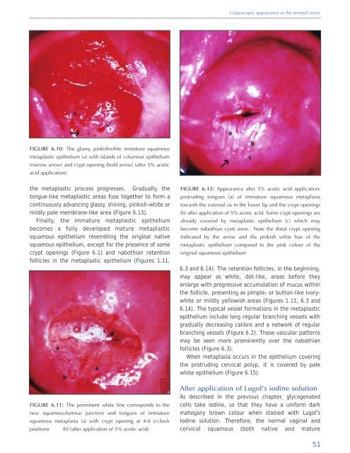
FIGURE 6.12: Appearance after 5% acetic acid application:<br />
protruding tongues (a) <strong>of</strong> immature squamous metaplasia<br />
towards the external os in the lower lip <strong>and</strong> the crypt openings<br />
(b) after application <strong>of</strong> 5% acetic acid. Some crypt openings are<br />
already covered by metaplastic epithelium (c) which may<br />
become nabothian cysts soon. Note the distal crypt opening<br />
indicated by the arrow <strong>and</strong> the pinkish white hue <strong>of</strong> the<br />
metaplastic epithelium compared to the pink colour <strong>of</strong> the<br />
original squamous epithelium<br />
6.3 <strong>and</strong> 6.14). The retention follicles, in the beginning,<br />
may appear as white, dot-like, areas before they<br />
enlarge with progressive accumulation <strong>of</strong> mucus within<br />
the follicle, presenting as pimple- or button-like ivorywhite<br />
or mildly yellowish areas (Figures 1.11, 6.3 <strong>and</strong><br />
6.14). The typical vessel formations in the metaplastic<br />
epithelium include long regular branching vessels with<br />
gradually decreasing calibre <strong>and</strong> a network <strong>of</strong> regular<br />
branching vessels (Figure 6.2). These vascular patterns<br />
may be seen more prominently over the nabothian<br />
follicles (Figure 6.3).<br />
When metaplasia occurs in the epithelium covering<br />
the protruding cervical polyp, it is covered by pale<br />
white epithelium (Figure 6.15).<br />
After application <strong>of</strong> Lugol’s iodine solution<br />
As described in the previous chapter, glycogenated<br />
cells take iodine, so that they have a uniform dark<br />
mahogany brown colour when stained with Lugol’s<br />
iodine solution. Therefore, the normal vaginal <strong>and</strong><br />
cervical squamous (both native <strong>and</strong> mature<br />
51


