

Colposcopy and Treatment of Cervical Intraepithelial Neoplasia - RHO
Colposcopy and Treatment of Cervical Intraepithelial Neoplasia - RHO
Colposcopy and Treatment of Cervical Intraepithelial Neoplasia - RHO

You also want an ePaper? Increase the reach of your titles
YUMPU automatically turns print PDFs into web optimized ePapers that Google loves.
Chapter 2<br />
cytoplasm in relation to the size <strong>of</strong> the nucleus<br />
(nuclear-cytoplasmic ratio) is one <strong>of</strong> the most<br />
important base for assessing the grade <strong>of</strong> CIN (Figure<br />
2.1). Increased ratios are associated with more severe<br />
degrees <strong>of</strong> CIN. More <strong>of</strong>ten than not, a cervical smear<br />
contains cells with a range <strong>of</strong> changes; considerable<br />
challenges <strong>and</strong> subjectivity, therefore, are involved in<br />
reporting the results. Experience <strong>of</strong> the cytologist is<br />
critically important in final reporting.<br />
Diagnosis <strong>and</strong> grading <strong>of</strong> CIN by<br />
histopathology<br />
CIN may be suspected through cytological examination<br />
using the Papanicolaou technique or through<br />
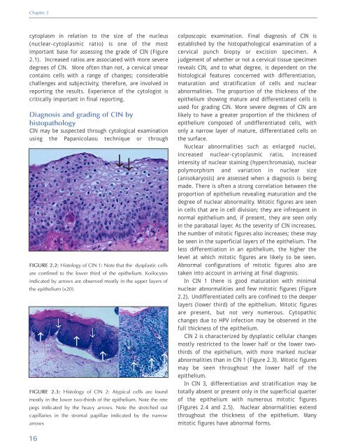
FIGURE 2.2: Histology <strong>of</strong> CIN 1: Note that the dysplastic cells<br />
are confined to the lower third <strong>of</strong> the epithelium. Koilocytes<br />
indicated by arrows are observed mostly in the upper layers <strong>of</strong><br />
the epithelium (x20).<br />
↑<br />
↓<br />
↑<br />
↓<br />
↑<br />
↓ ↓<br />
FIGURE 2.3: Histology <strong>of</strong> CIN 2: Atypical cells are found<br />
mostly in the lower two-thirds <strong>of</strong> the epithelium. Note the rete<br />
pegs indicated by the heavy arrows. Note the stretched out<br />
capillaries in the stromal papillae indicated by the narrow<br />
arrows<br />
↓<br />
↑<br />
↓<br />
↑ ↑<br />
colposcopic examination. Final diagnosis <strong>of</strong> CIN is<br />
established by the histopathological examination <strong>of</strong> a<br />
cervical punch biopsy or excision specimen. A<br />
judgement <strong>of</strong> whether or not a cervical tissue specimen<br />
reveals CIN, <strong>and</strong> to what degree, is dependent on the<br />
histological features concerned with differentiation,<br />
maturation <strong>and</strong> stratification <strong>of</strong> cells <strong>and</strong> nuclear<br />
abnormalities. The proportion <strong>of</strong> the thickness <strong>of</strong> the<br />
epithelium showing mature <strong>and</strong> differentiated cells is<br />
used for grading CIN. More severe degrees <strong>of</strong> CIN are<br />
likely to have a greater proportion <strong>of</strong> the thickness <strong>of</strong><br />
epithelium composed <strong>of</strong> undifferentiated cells, with<br />
only a narrow layer <strong>of</strong> mature, differentiated cells on<br />
the surface.<br />
Nuclear abnormalities such as enlarged nuclei,<br />
increased nuclear-cytoplasmic ratio, increased<br />
intensity <strong>of</strong> nuclear staining (hyperchromasia), nuclear<br />
polymorphism <strong>and</strong> variation in nuclear size<br />
(anisokaryosis) are assessed when a diagnosis is being<br />
made. There is <strong>of</strong>ten a strong correlation between the<br />
proportion <strong>of</strong> epithelium revealing maturation <strong>and</strong> the<br />
degree <strong>of</strong> nuclear abnormality. Mitotic figures are seen<br />
in cells that are in cell division; they are infrequent in<br />
normal epithelium <strong>and</strong>, if present, they are seen only<br />
in the parabasal layer. As the severity <strong>of</strong> CIN increases,<br />
the number <strong>of</strong> mitotic figures also increases; these may<br />
be seen in the superficial layers <strong>of</strong> the epithelium. The<br />
less differentiation in an epithelium, the higher the<br />
level at which mitotic figures are likely to be seen.<br />
Abnormal configurations <strong>of</strong> mitotic figures also are<br />
taken into account in arriving at final diagnosis.<br />
In CIN 1 there is good maturation with minimal<br />
nuclear abnormalities <strong>and</strong> few mitotic figures (Figure<br />
2.2). Undifferentiated cells are confined to the deeper<br />
layers (lower third) <strong>of</strong> the epithelium. Mitotic figures<br />
are present, but not very numerous. Cytopathic<br />
changes due to HPV infection may be observed in the<br />
full thickness <strong>of</strong> the epithelium.<br />
CIN 2 is characterized by dysplastic cellular changes<br />
mostly restricted to the lower half or the lower twothirds<br />
<strong>of</strong> the epithelium, with more marked nuclear<br />
abnormalities than in CIN 1 (Figure 2.3). Mitotic figures<br />
may be seen throughout the lower half <strong>of</strong> the<br />
epithelium.<br />
In CIN 3, differentiation <strong>and</strong> stratification may be<br />
totally absent or present only in the superficial quarter<br />
<strong>of</strong> the epithelium with numerous mitotic figures<br />
(Figures 2.4 <strong>and</strong> 2.5). Nuclear abnormalities extend<br />
throughout the thickness <strong>of</strong> the epithelium. Many<br />
mitotic figures have abnormal forms.<br />
16


