

Colposcopy and Treatment of Cervical Intraepithelial Neoplasia - RHO
Colposcopy and Treatment of Cervical Intraepithelial Neoplasia - RHO
Colposcopy and Treatment of Cervical Intraepithelial Neoplasia - RHO

Create successful ePaper yourself
Turn your PDF publications into a flip-book with our unique Google optimized e-Paper software.
Colposcopic assessment <strong>of</strong> cervical intraepithelial neoplasia<br />
harbour atypical vessels; CIN 3 lesions tend to be<br />
complex, involving the os.<br />
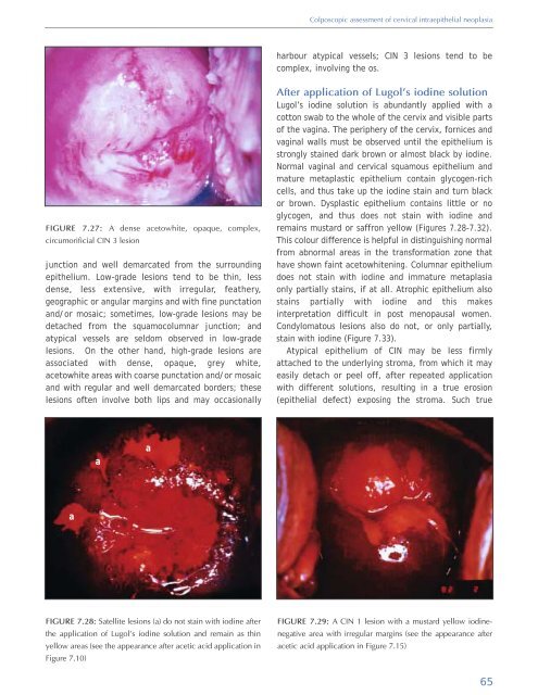
FIGURE 7.27: A dense acetowhite, opaque, complex,<br />
circumorificial CIN 3 lesion<br />
junction <strong>and</strong> well demarcated from the surrounding<br />
epithelium. Low-grade lesions tend to be thin, less<br />
dense, less extensive, with irregular, feathery,<br />
geographic or angular margins <strong>and</strong> with fine punctation<br />
<strong>and</strong>/or mosaic; sometimes, low-grade lesions may be<br />
detached from the squamocolumnar junction; <strong>and</strong><br />
atypical vessels are seldom observed in low-grade<br />
lesions. On the other h<strong>and</strong>, high-grade lesions are<br />
associated with dense, opaque, grey white,<br />
acetowhite areas with coarse punctation <strong>and</strong>/or mosaic<br />
<strong>and</strong> with regular <strong>and</strong> well demarcated borders; these<br />
lesions <strong>of</strong>ten involve both lips <strong>and</strong> may occasionally<br />
After application <strong>of</strong> Lugol’s iodine solution<br />
Lugol’s iodine solution is abundantly applied with a<br />
cotton swab to the whole <strong>of</strong> the cervix <strong>and</strong> visible parts<br />
<strong>of</strong> the vagina. The periphery <strong>of</strong> the cervix, fornices <strong>and</strong><br />
vaginal walls must be observed until the epithelium is<br />
strongly stained dark brown or almost black by iodine.<br />
Normal vaginal <strong>and</strong> cervical squamous epithelium <strong>and</strong><br />
mature metaplastic epithelium contain glycogen-rich<br />
cells, <strong>and</strong> thus take up the iodine stain <strong>and</strong> turn black<br />
or brown. Dysplastic epithelium contains little or no<br />
glycogen, <strong>and</strong> thus does not stain with iodine <strong>and</strong><br />
remains mustard or saffron yellow (Figures 7.28-7.32).<br />
This colour difference is helpful in distinguishing normal<br />
from abnormal areas in the transformation zone that<br />
have shown faint acetowhitening. Columnar epithelium<br />
does not stain with iodine <strong>and</strong> immature metaplasia<br />
only partially stains, if at all. Atrophic epithelium also<br />
stains partially with iodine <strong>and</strong> this makes<br />
interpretation difficult in post menopausal women.<br />
Condylomatous lesions also do not, or only partially,<br />
stain with iodine (Figure 7.33).<br />
Atypical epithelium <strong>of</strong> CIN may be less firmly<br />
attached to the underlying stroma, from which it may<br />
easily detach or peel <strong>of</strong>f, after repeated application<br />
with different solutions, resulting in a true erosion<br />
(epithelial defect) exposing the stroma. Such true<br />
a<br />
a<br />
a<br />
FIGURE 7.28: Satellite lesions (a) do not stain with iodine after<br />
the application <strong>of</strong> Lugol’s iodine solution <strong>and</strong> remain as thin<br />
yellow areas (see the appearance after acetic acid application in<br />
Figure 7.10)<br />
FIGURE 7.29: A CIN 1 lesion with a mustard yellow iodinenegative<br />
area with irregular margins (see the appearance after<br />
acetic acid application in Figure 7.15)<br />
65


