

Colposcopy and Treatment of Cervical Intraepithelial Neoplasia - RHO
Colposcopy and Treatment of Cervical Intraepithelial Neoplasia - RHO
Colposcopy and Treatment of Cervical Intraepithelial Neoplasia - RHO

Create successful ePaper yourself
Turn your PDF publications into a flip-book with our unique Google optimized e-Paper software.
An introduction to cervical intraepithelial neoplasia (CIN)<br />
Table 2.4: Natural history <strong>of</strong> SIL<br />
Baseline cytological<br />
abnormality<br />
Regression to normal<br />
at 24 months<br />
Progression to HSIL at<br />
24 months<br />
Progression to invasive<br />
cancer at 24 months<br />
ASCUS<br />
LSIL<br />
HSIL<br />
68.2%<br />
47.4%<br />
35.0%<br />
7.1%<br />
20.8%<br />
23.4% (persistence)<br />
0.3%<br />
0.2%<br />
1.4%<br />
abnormality given in Table 2.4 (Melinkow et al., 1998).<br />
Holowaty et al., (1999) calculated RR <strong>of</strong> progression<br />
<strong>and</strong> regression by 2-years <strong>of</strong> follow-up for moderate<br />
<strong>and</strong> severe dysplasias, with mild dysplasia taken as the<br />
baseline reference category. RRs for CIS were 8.1 for<br />
moderate dysplasia <strong>and</strong> 22.7 for severe dysplasia. The<br />
corresponding RRs for invasive cancer were 4.5 <strong>and</strong><br />
20.7, respectively.<br />
Adenocarcinoma in situ<br />
The precursor lesion that has been recognized to arise<br />
from the columnar epithelium is referred to as<br />
adenocarcinoma in situ (AIS). In AIS, normal columnar<br />
epithelium is replaced by abnormal epithelium<br />
showing loss <strong>of</strong> polarity, increased cell size, increased<br />
nuclear size, nuclear hyperchromasia, mitotic activity,<br />
reduction <strong>of</strong> cytoplasmic mucin expression <strong>and</strong> cellular<br />
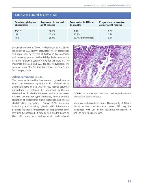
stratification or piling (Figure 2.6). Abnormal<br />
branching <strong>and</strong> budding gl<strong>and</strong>s with intraluminal<br />
papillary epithelial projections lacking stromal cores<br />
may also be observed. It may be sub-divided based on<br />
the cell types into endocervical, endometroid,<br />
FIGURE 2.6: Adenocarcinoma in situ, coexisting with a normal<br />
endocervical epithelium (x10).<br />
intestinal <strong>and</strong> mixed cell types. The majority <strong>of</strong> AIS are<br />
found in the transformation zone. AIS may be<br />
associated with CIN <strong>of</strong> the squamous epithelium in<br />
one- to two-thirds <strong>of</strong> cases.<br />
19


