

Colposcopy and Treatment of Cervical Intraepithelial Neoplasia - RHO
Colposcopy and Treatment of Cervical Intraepithelial Neoplasia - RHO
Colposcopy and Treatment of Cervical Intraepithelial Neoplasia - RHO

You also want an ePaper? Increase the reach of your titles
YUMPU automatically turns print PDFs into web optimized ePapers that Google loves.
Chapter 4<br />
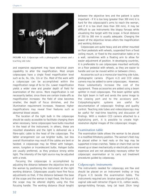
FIGURE 4.2: Colposcope with a photographic camera <strong>and</strong> a<br />
teaching side tube<br />
<strong>and</strong> expensive equipment may have electrical zoom<br />
capability to alter the magnification. Most simple<br />
colposcopes have a single fixed magnification level<br />
such as 6x, 9x, 10x, 12x or 15x. Most <strong>of</strong> the work with<br />
a colposcope can be accomplished within the<br />
magnification range <strong>of</strong> 6x to 15x. Lower magnification<br />
yields a wider view <strong>and</strong> greater depth <strong>of</strong> field for<br />
examination <strong>of</strong> the cervix. More magnification is not<br />
necessarily better, since there are certain trade-<strong>of</strong>fs as<br />
magnification increases: the field <strong>of</strong> view becomes<br />
smaller, the depth <strong>of</strong> focus dimishes, <strong>and</strong> the<br />
illumination requirement increases. However, higher<br />
magnifications may reveal finer features such as<br />
abnormal blood vessels.<br />
The location <strong>of</strong> the light bulb in the colposcope<br />
should be easily accessible to facilitate changing them<br />
when necessary. Some colposcopes have bulbs mounted<br />
in the head <strong>of</strong> the instrument; in others, these are<br />
mounted elsewhere <strong>and</strong> the light is delivered via a<br />
fibre-optic cable to the head <strong>of</strong> the colposcope. The<br />
latter arrangement can use brighter bulbs, but less<br />
overall illumination may result if the cables are bent or<br />
twisted. A colposcope may be fitted with halogen,<br />
xenon, tungsten or inc<strong>and</strong>escent bulbs. Halogen bulbs<br />
are usually preferred, as they produce strong white<br />
light. The intensity <strong>of</strong> the light source may be adjusted<br />
with a knob.<br />
Focusing the colposcope is accomplished by<br />
adjusting the distance between the objective lens <strong>and</strong><br />
the woman by positioning the instrument at the right<br />
working distance. Colposcopes usually have fine focus<br />
adjustments so that, if the distance between the base<br />
<strong>of</strong> the scope <strong>and</strong> the woman is kept fixed, the focus <strong>of</strong><br />
the scope may be altered slightly using the fine<br />
focusing h<strong>and</strong>le. The working distance (focal length)<br />
between the objective lens <strong>and</strong> the patient is quite<br />
important - if it is too long (greater than 300 mm) it is<br />
hard for the colposcopist’s arms to reach the woman,<br />
<strong>and</strong> if it is too short (less than 200 mm), it may be<br />
difficult to use instruments like biopsy forceps while<br />
visualizing the target with the scope. A focal distance<br />
<strong>of</strong> 250 to 300 mm is usually adequate. Changing the<br />
power <strong>of</strong> the objective lenses alters the magnification<br />
<strong>and</strong> working distance.<br />
Colposcopes are quite heavy <strong>and</strong> are either mounted<br />
on floor pedestals with wheels, suspended from a fixed<br />
ceiling mount, or fixed to the examination table or to<br />
a wall, sometimes with a floating arm to allow for<br />
easier adjustment <strong>of</strong> position. In developing countries,<br />
it is preferable to use colposcopes mounted vertically<br />
on a floor pedestal with wheels, as they are easier to<br />
h<strong>and</strong>le <strong>and</strong> can be moved within or between clinics.<br />
Accessories such as a monocular teaching side tube,<br />
photographic camera (Figure 4.2) <strong>and</strong> CCD video<br />
camera may be added to some colposcopes. However,<br />
these substantially increase the cost <strong>of</strong> the<br />
equipment. These accessories are added using a beam<br />
splitter in most colposcopes. The beam splitter splits<br />
the light beam in half <strong>and</strong> sends the same image to<br />
the viewing port <strong>and</strong> to the accessory port.<br />
Colpophotographic systems are useful for<br />
documentation <strong>of</strong> colposcopic findings <strong>and</strong> quality<br />
control. Teaching side tubes <strong>and</strong> videocolposcopy may<br />
be useful for real-time teaching <strong>and</strong> discussion <strong>of</strong><br />
findings. With a modern CCD camera attached to a<br />
digitalizing port, it is possible to create highresolution<br />
digital images <strong>of</strong> the colposcopic images.<br />
Examination table<br />
The examination table allows the woman to be placed<br />
in a modified lithotomy position. The woman’s feet may<br />
be placed either in heel rests or the legs may be<br />
supported in knee crutches. Tables or chairs that can be<br />
moved up or down mechanically or electrically are more<br />
expensive <strong>and</strong> are not absolutely necessary either for<br />
colposcopic examination or to carry out treatment<br />
procedures guided by colposcopy.<br />
Colposcopic instruments<br />
The instruments needed for colposcopy are few <strong>and</strong><br />
should be placed on an instrument trolley or tray<br />
(Figure 4.3) beside the examination table. The<br />
instruments required are: bivalve specula (Figure 4.4),<br />
vaginal side-wall retractor (Figure 4.5), cotton swabs,<br />
sponge-holding forceps, long (at least 20cm long)<br />
32


